MRI Testis
Indications for testicular mri scn
- Characterization of testicular neoplasms
- Hydrocele, spermatocele, and varicocele
- Testicular epidermoid cyst
- Looking for ectopic testis
- Segmental testicular infarction
- Epididymitis and orchitis
- Testicular lumps
- Testicular cancer
- Dystopic testis
- Trauma
Contraindications for MRI Testis
- Any electrically, magnetically or mechanically activated implant (e.g. cardiac pacemaker, insulin pump biostimulator, neurostimulator, cochlear implant, and hearing aids)
- Intracranial aneurysm clips (unless made of titanium)
- Pregnancy (risk vs benefit ratio to be assessed)
- Ferromagnetic surgical clips or staples
- Metallic foreign body in the eye
- Metal shrapnel or bullet
Patient preparation for MRI Testis
- A satisfactory written consent form must be taken from the patient before entering the scanner room
- Ask the patient to remove all metal objects including keys, coins, wallet, cards with magnetic strips, jewellery, hearing aid and hairpins
- Ask the patient to undress and change into a hospital gown
- Contrast injection risk and benefits must be explained to the before the scan
- Gadolinium should only be given to the patient if GFR is > 30
- If possible provide a chaperone for claustrophobic patients (e.g. relative or staff )
- Offer earplugs or headphones, possibly with music for extra comfort
- Explain the procedure to the patient
- Instruct the patient to keep still
- Note the weight of the patient
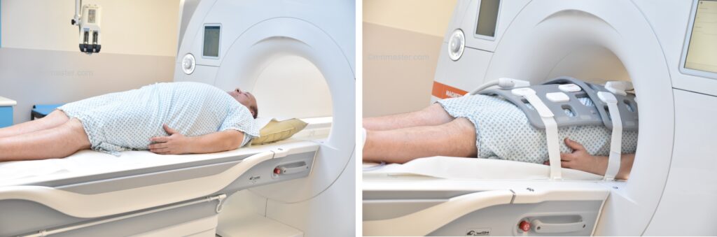
Positioning for MRI Testis
- Position the patient in supine position with head pointing towards the magnet (head first supine)
- Position the patient over the spine coil and place the body coil over the pelvis( iliac crest down to three inches below testicle)
- Securely tighten the body coil using straps to prevent respiratory artefacts
- Give a pillow under the head for extra comfort (do not give cushions under the legs )
- Centre the laser beam localiser over pubic symphysis
Recommended MRI Testis Protocols and Planning
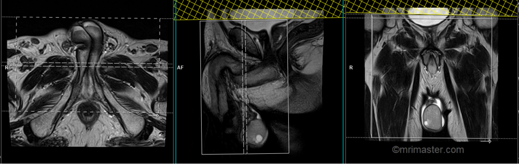
MRI Testis localiser
A three-plane HASTE localizer must be taken initially to localize and plan the sequences. These are fast single-shot localizers with under 25s acquisition time, which are excellent for localizing abdominal and pelvic structures. Take at least 3-4 slices in all planes to get the best results.

T2 stir coronal 5 mm big FOV
Plan the big FOV coronal slices on the sagittal plane, angling the positioning block vertically across the abdomen. Check the positioning block in the other two planes. An appropriate angle must be given in the axial plane (parallel to the right and left hip joint). The slices must be sufficient to cover the abdomen and pelvis from the anterior abdominal wall to the spinous process of the vertebrae.
The FOV must be sufficiently large to encompass the abdomen and pelvis from the kidneys down to the scrotum, typically ranging from 400mm to 420mm. Large FOV scans are essential for examining ectopic testis, as the testicles can be located anywhere along the gonadal vein. Therefore, it is crucial to cover the region from the kidneys down to the scrotum. Additionally, these scans are conducted to assess the para-aortic and pre-sacral nodes in testicular cancer imaging.

Parameters
TR 4000-5000 | TE 110 | FLIP 130 | NEX 2 | SLICE 5MM | MATRIX 384X384 | FOV 400-420 | PHASE R>L | GAP 10% | TI 150 |
T2 tse\HASTE axial 5 mm big FOV
Plan the big FOV axial slices on the coronal plane. Angle the positioning block parallel to the right and left iliac crest). Check the positioning block in the other two planes. An appropriate angle must be given in the sagittal plane (horizontally across the abdomen). Slices must be sufficient to cover the lower abdomen and pelvis from the middle of the kidneys down to the scrotum.
For the big FOV scans, it is essential to ensure sufficient coverage of the entire pelvis, typically ranging from 350mm to 400mm. These extensive scans are particularly useful in checking for ectopic testis, as the testicles can be present anywhere along the gonadal vein. Therefore, it is vital to encompass the area from the kidneys down to the scrotum to effectively detect any abnormalities related to ectopic testis.

Parameters
TR 4000-5000 | TE 100-120 | SLICE 5 MM | FLIP 130-150 | PHASE A>P | MATRIX 512X384 | FOV 350-400 | GAP 10% | NEX(AVRAGE) 2 |
T2 tse sagittal 3mm SFOV
Plan the sagittal slices on the axial plane. Angle the positioning block parallel to the penile shaft and the anal canal. Check the positioning block in the other two planes. An appropriate angle must be given in the coronal plane, parallel to the intrapubic fibrocartilage. Slices must be sufficient to cover the entire scrotum from the right acetabulum to the left acetabulum. The field of view (FOV) must be big enough to cover the scrotum and prostate (normally 200mm-250mm).

Parameters
TR 4000-5000 | TE 100-120 | SLICE 3 MM | FLIP 130-150 | PHASE H>F | MATRIX 320X320 | FOV 250-300 | GAP 10% | NEX(AVRAGE) 3 |
T2 stir axial 3mm SFOV
Plan the axial slices on the sagittal plane; angle the positioning block horizontally across the scrotum (parallel to the penile shaft). Check the positioning block in the other two planes. Ensure an appropriate angle is given in the coronal plane (horizontally across the testis). The slices must be sufficient to cover the scrotum from the prostate down to 1 cm below the scrotum.
Parameters

TR 4000-5000 | TE 110 | FLIP 150 | NEX 4 | SLICE 3 MM | MATRIX 256X256 | FOV 180-200 | PHASE R>L | GAP 10% | TI 150 |
T1 tse axial 3mm SFOV
Plan the axial slices on the sagittal plane; angle the positioning block horizontally across the scrotum (parallel to the penile shaft). Check the positioning block in the other two planes. Ensure an appropriate angle is given in the coronal plane (horizontally across the testis). The slices must be sufficient to cover the scrotum from the prostate down to 1 cm below the scrotum.
Parameters
TR 500-600 | TE 15-25 | SLICE 3 MM | FLIP 140 | PHASE R>L | MATRIX 320X256 | FOV 180-200 | GAP 10% | NEX(AVRAGE) 3 |
T2 tse axial 3mm SFOV
Plan the axial slices on the sagittal plane; angle the positioning block horizontally across the scrotum (parallel to the penile shaft). Check the positioning block in the other two planes. Ensure an appropriate angle is given in the coronal plane (horizontally across the testis). The slices must be sufficient to cover the scrotum from the prostate down to 1 cm below the scrotum.
Parameters
TR 4000-5000 | TE 100-120 | SLICE 3 MM | FLIP 130-150 | PHASE R>L | MATRIX 320X256 | FOV 180-230 | GAP 10% | NEX(AVRAGE) 3 |
T2 tse coronal 3mm SFOV
Plan the coronal slices on the axial plane and angle the positioning block perpendicular to the penile shaft. Check the positioning block in the other two planes. An appropriate angle must be given in the sagittal plane (vertically across the scrotum). Slices must be sufficient to cover the entire scrotum from the penis to the anal canal.
Parameters
TR 4000-5000 | TE 100-120 | SLICE 3 MM | FLIP 130-150 | PHASE R>L | MATRIX 320X256 | FOV 180-230 | GAP 10% | NEX(AVRAGE) 4 |
T1 tse coronal 3mm SFOV
Plan the coronal slices on the axial plane and angle the positioning block perpendicular to the penile shaft. Check the positioning block in the other two planes. An appropriate angle must be given in the sagittal plane (vertically across the scrotum). Slices must be sufficient to cover the entire scrotum from the penis to the anal canal.
Parameters
TR 400-600 | TE 15-25 | SLICE 3 MM | FLIP 140 | PHASE R>L | MATRIX 320X256 | FOV 180-200 | GAP 10% | NEX(AVRAGE) 4 |
Scrotum scans rarely require contrast-enhanced imaging. However, when contrast-enhanced imaging is necessary, T1 TSE Fat-saturated small FOV axial and coronal scans should be performed following the administration of gadolinium injection (using the same planning as described earlier).