Appendix MRI Protocol and Planning
Indications for Appendix mri
- Right iliac fossa (RIF) pain
- inflammatory bowel disease
- appendicitis
Contraindications
- Any electrically, magnetically or mechanically activated implant (e.g. cardiac pacemaker, insulin pump biostimulator, neurostimulator, cochlear implant, and hearing aids)
- Intracranial aneurysm clips (unless made of titanium)
- Pregnancy (risk vs benefit ratio to be assessed)
- Ferromagnetic surgical clips or staples
- Metallic foreign body in the eye
- Metal shrapnel or bullet
Patient preparation for Appendix MRI
- A satisfactory written consent form must be taken from the patient before entering the scanner room
- Ask the patient to remove all metal objects including keys, coins, wallet, cards with magnetic strips, jewellery, hearing aid and hairpins
- Ask the patient to undress and change into a hospital gown
- Instruct the patient to hold their breath for the breath hold scans and breathe gently for the gated scans. It is recommended to provide coaching to the patient two to three times before initiating the scan.
- Claustrophobic patients may be accompanied into the scanner room e.g. by staff member or relative with proper safety screening
- Buscopan injection risk and benefits must be explained to the patient before the scan
- Offer headphones for communicating with the patient and ear protection
- Explain the procedure to the patient and answer questions
- Note down the weight of the patient
Positioning for Appendix MRI
- Position the patient in supine position with head pointing towards the magnet (head first supine)
- Position the patient over the spine coil and position the body coil over the lower abdomen and pelvis (from the xiphisternum down to the symphysis pubis).
- Securely tighten the body coil using straps to prevent respiratory artefacts
- Give a pillow under the head for extra comfort (do not give cushions under the legs )
- Centre the laser beam localiser over iliac crest

Recommended Appendix MRI Protocols and Planning
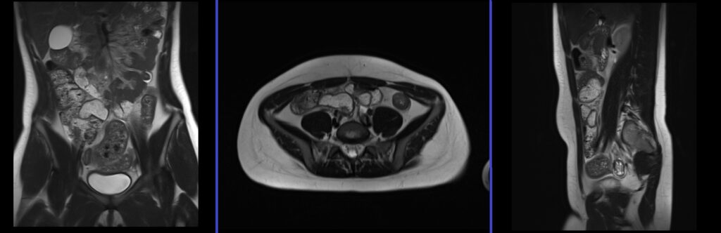
Appendix MRILlocaliser
To localize and plan the sequences, it is essential to acquire a three-plane T2 HASTE localizer initially. These fast single-shot localizers have an acquisition time of under 25 seconds and are highly effective in accurately localizing abdominal structures.
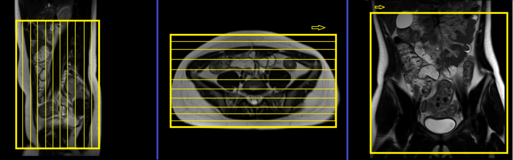
T2 HASTE 5 mm breath hold coronal
Plan the coronal slices based on the axial image, positioning the block horizontally across the abdomen as shown below. Confirm the positioning in the other two planes. Establish an appropriate angle in the sagittal plane, aligning it vertically across the abdomen. Ensure that an adequate number of slices are acquired to cover the abdomen and pelvis, extending from the anterior abdominal wall to the spinal canal. The field of view (FOV) should be sufficiently large to encompass the abdomen and pelvis, from the xiphisternum to the pubic symphysis. To prevent wrap-around artifacts, employ phase oversampling. Instruct the patient to hold their breath during image acquisition.
Parameters
TR 2000-2500 | TE 90-110 | FLIP 130 | NEX 1 | SLICE 5MM | MATRIX 256×256 | FOV 350 | PHASE R>L | OVERSAMPLE 50% | TRIGGER NO |
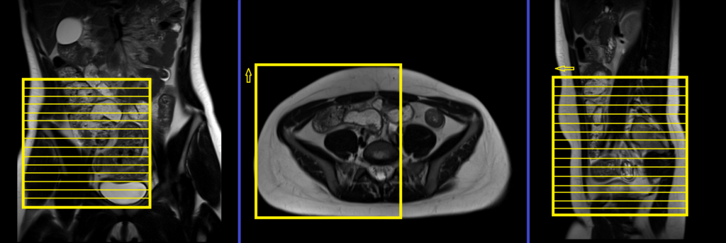
T2 HASTE 5 mm breath hold axial
Plan the axial slices on the coronal image by positioning the block horizontally across the abdomen as shown below. Verify the positioning in the other two planes. Establish an appropriate angle in the sagittal plane, aligning it horizontally across the abdomen. Ensure that the slices are adequate to cover the abdomen and pelvis, extending from the hepatic flexure of the colon superiorly to the pubic symphysis inferiorly. Phase oversampling can be employed to prevent wrap-around artifacts. Instruct the patient to hold their breath during image acquisition.

TR 2500-3000 | TE 90-110 | FLIP 130 | NEX 1 | SLICE 5MM | MATRIX 256×256 | FOV 350 | PHASE R>L | OVERSAMPLE 50% | TRIGGER NO |
Pause for buscopan injection
Warning
* Buscopan injection should not be administered to patients with myasthenia gravis, megacolon, narrow angle glaucoma, tachycardia, prostatic enlargement with urinary retention, mechanical stenoses in the region of the gastrointestinal tract or paralytic ileus.*
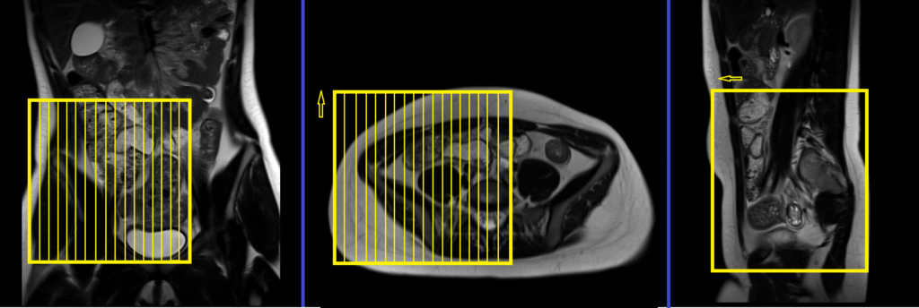
T2 TSE axial multiple breath holds 4mm SFOV
Plan the axial slices on the coronal image. Angle the positioning block across the ascending colon and cecum as demonstrated below. Ensure that the positioning block is checked in the other two planes. Establish an appropriate angle in the sagittal plane perpendicular to the rectus abdominis muscle. The slices should adequately cover the right iliac fossa, spanning from the mid ascending colon superiorly to the pubic symphysis inferiorly. Select a sufficient SFOV (normally 200mm-250mm) to encompass the affected area. To prevent wrap-around artifacts, employ phase oversampling. Additionally, adding saturation bands at the top and bottom of the axial block will minimize arterial pulsation and breathing artifacts. Instruct the patient to hold their breath during image acquisition.
This sequence is a modified version of a T2 TSE breath-hold scan commonly used in abdominal and liver imaging. To obtain high-resolution breath-hold scans, users can customize the T2 sequence. The default sequence parameters are as follows: 350-400 FOV, matrix 320×320, NEX 1, slice thickness 6mm, and acquisition of 25-30 slices within a 30-second breath hold. To achieve the desired high-resolution scan, make the following modifications: 200-250 FOV, matrix 256×204, NEX 2, and a slice thickness of 4mm with parallel imaging (IPAT) enabled. Typically, the adapted sequence will take approximately 90 seconds, requiring it to be divided into 4 acquisitions (concatenations). This division ensures that each breath-hold acquisition lasts approximately 22 seconds.
Parameters
TR 3000-4000 | TE 100 | FLIP 150 | NEX 2 | SLICE 4 MM | MATRIX 256X192 | FOV 200-250 | PHASE A>P | OVERSAMPLE 30% | IPAT On |
T1 VIBE DIXON SFOV 4mm axial breath hold
Plan the axial slices on the coronal image. Angle the positioning block across the ascending colon and cecum as demonstrated below. Ensure that the positioning block is checked in the other two planes. Establish an appropriate angle in the sagittal plane perpendicular to the rectus abdominis muscle. The slices should adequately cover the right iliac fossa, spanning from the mid ascending colon superiorly to the pubic symphysis inferiorly. Select a sufficient SFOV (normally 200mm-250mm) to encompass the affected area. To prevent wrap-around artifacts, employ phase oversampling. Additionally, adding saturation bands at the top and bottom of the axial block will minimize arterial pulsation and breathing artifacts. Instruct the patient to hold their breath during image acquisition.
Parameters
T2 TSE fat sat/ STIR axial multiple breath holds 4mm SFOV
Plan the axial slices on the coronal image. Angle the positioning block across the ascending colon and cecum as demonstrated below. Ensure that the positioning block is checked in the other two planes. Establish an appropriate angle in the sagittal plane perpendicular to the rectus abdominis muscle. The slices should adequately cover the right iliac fossa, spanning from the mid ascending colon superiorly to the pubic symphysis inferiorly. Select a sufficient SFOV (normally 200mm-250mm) to encompass the affected area. To prevent wrap-around artifacts, employ phase oversampling. Additionally, adding saturation bands at the top and bottom of the axial block will minimize arterial pulsation and breathing artifacts. Instruct the patient to hold their breath during image acquisition.
Parameters
TR 4000-5000 | TE 500 | FLIP 150 | NEX 1 | SLICE 40MM | MATRIX 320X320 | FOV 350-450 | PHASE R>L | OVERSAMPLE 50% | IPAT ON |
T2 TSE coronal multiple breath holds 4mm SFOV
Plan the coronal slices on the axial image, angling the positioning block horizontally across the abdomen as depicted below. Confirm the positioning in the other two planes. Establish an appropriate angle in the sagittal plane parallel to the rectus abdominis muscle. The slices should adequately cover the right iliac fossa from the anterior abdominal wall to the spinal canal. Select a small field of view (FOV) that is sufficiently large to encompass the affected area (typically 200mm-250mm). To prevent wrap-around artifacts, employ phase oversampling. Instruct the patient to hold their breath during image acquisition.

Parameters
TR 3000-4000 | TE 100 | FLIP 150 | NEX 2 | SLICE 4 MM | MATRIX 256X192 | FOV 200-250 | PHASE R>L | OVERSAMPLE 70% | IPAT On |
T2 TSE sagittal multiple breath holds 4mm SFOV
Plan the sagittal slices on the axial image, angling the positioning block parallel to the ascending colon and cecum. Verify the positioning block in the other two planes. Ensure an appropriate angle is maintained in the axial plane, approximately perpendicular to the rectus abdominis muscle. The slices should adequately cover the right iliac fossa and hypogastric region. Select a small field of view (FOV) that adequately encompasses the affected area (typically 200mm-250mm). Utilize phase oversampling to prevent wrap-around artifacts. Instruct the patient to hold their breath during image acquisition.
Parameters
TR 3000-4000 | TE 100 | FLIP 150 | NEX 2 | SLICE 4 MM | MATRIX 256X192 | FOV 200-250 | PHASE A>P | OVERSAMPLE 30% | IPAT On |