T1 Fat Suppressed MRI Sequence
Fat saturation is an MRI technique used to suppress the signal from normal adipose tissue to reduce chemical shift artefact, improve visualization of uptake of contrast material and tissue characterization. To suppress the fat signal from an MRI sequence, a special fat suppression module is inserted at the beginning of a normal MRI sequence. Fat suppression in an MRI sequence can be achieved with five techniques: spectral fat saturation, short tau inversion recovery (STIR), spectral presaturation with inversion recovery (SPAIR), dixon method and water excitation method.
MRI image appearance of T1 fat suppressed MRI
The easiest way to identify T1 weighted fat saturated images is to look for adipose tissues in the body (e.g. subcutaneous fat and fat in bone marrow). Areas contain adipose tissues appear dark on T1 weighted fat saturated images. All the other characteristics of the T1 weighted fat saturated images remain the same as the T1 weighted images. Here’s how different tissues appear on T1 fat-saturated MRI:
- Fat Tissue: Fat appears dark on T1 fat-saturated MRI due to the suppression of its signal.
- Muscle Tissue: Muscles typically appear with intermediate signal intensity(gray) on T1 fat-saturated MRI.
- Liver: The normal liver tissue appears intermediate in signal intensity, similar to muscle.
- Kidney: Normal kidney tissue typically appears as intermediate in signal intensity on T1-fat saturated images.
- Pancreas: The pancreas can have variable signal intensity, but it’s generally intermediate.
- Spleen: The spleen typically has a signal intensity similar to the liver or muscle, being intermediate on T1-fat saturated images.
- Cerebrospinal Fluid (CSF) and Synovial Fluid: CSF and synovial fluid both appear dark (hypointense) on T1 fat-saturated MRI.
- Urinary Bladder: The urinary bladder, when filled with urine, may appear with intermediate signal intensity. The appearance can vary depending on the amount of fluid present.
- Bile Duct: The common bile duct is usually not well visualized on T1 fat-saturated MRI because it contains a mixture of fluids, bile, and surrounding tissues. It may appear as a low-intensity structure in the right context.
- Bone: Bone appears dark on T1 fat-saturated MRI.
Pathological appearance on T1 fat-saturated MRI
- Pathologies containing fat: Pathologies containing adipose tissue will appear dark on T1-weighted fat-saturated images (e.g., lipoma).
- Solid Tumors: Solid tumors often appear hypointense (darker) or iso-intense on T1 fat-saturated images compared to surrounding healthy tissue. The degree of darkness can vary depending on the tumor’s composition and vascularity.
- Cystic Tumors: If a tumor contains cystic components, the cysts themselves may appear hypointense (darker) due to the fluid content within them, while the solid parts of the tumor remain hyperintense.
- Cysts: Simple fluid-filled cysts typically appear hypointense (darker) on T1 fat-saturated images due to the high water content within the cyst.
- Infarctions: Infarctions, which result from a lack of blood supply to a tissue, can appear hypointense on T1 fat-saturated images.
- Hemorrhages:
Acute Hemorrhages: Acute hemorrhages, such as those from a recent injury or bleeding event, often appear hypointense (darker) on T1 fat-saturated images due to the conversion of deoxyhemoglobin to intracellular methemoglobin.
Chronic Hemorrhages: In chronic or older hemorrhages, the appearance may vary, and they could become isointense or even hypointense as the blood products break down and are metabolized.
- Infections: Infected tissues may have variable signal intensities; most often, they appear hypointense (darker) or iso-intense on T1 fat-saturated images.
Tissues and their T1 fat saturated appearance
Brain:
- CSF: Dark.
- Fat: Dark.
- White Matter: Intermediate.
- Gray Matter: Intermediate to dark.
- Bone (skull): Dark.
- Bone Marrow: Intermediate.
- Blood Vessels: Mostly Dark; Depending on flow characteristics, can be bright or dark
- Pituitary Gland: Intermediate.
- Choroid Plexus: Intermediate.
- Cerebellum: Gray matter Darker than white matter.
- Brain Stem: Intermediate.
- Sinuses: Dark (air-filled).
- Thalamus, Putamen, Hippocampus, Caudate Nucleus: Intermediate.
- Corpus Callosum: Intermediate.
- Pineal Gland: Intermediate.
Spine:
- Spinal Cord: Intermediate.
- CSF: Dark.
- Bone: Dark.
- Bone Marrow: Intermediate.
- Intervertebral Disc: Nucleus pulposus. and annulus are Intermediate to bright.
- Ligaments: Intermediate to Dark.
- Nerve Roots: Intermediate.
Abdomen and Pelvis:
- Liver: Intermediate.
- Gallbladder and Common Bile Duct: Dark
- Spleen: Intermediate.
- Kidney: Intermediate.
- Ureters: Intermediate.
- Pancreas: Intermediate.
- Urinary Bladder: Intermediate to dark.
- Prostate: Intermediate.
- Uterus: Intermediate.
Musculoskeletal:
- Muscle: Intermediate to dark
- Bone: Dark (low signal)
- Bone Marrow: Intermediate
- Blood Vessels: Mostly Dark; Depending on flow characteristics, can be bright or dark
- Fat: Dark
- Ligaments: Intermediate to dark
- Nerve Roots: Intermediate
- Cartilage: Intermediate
- Synovial Fluid: Dark
- Tendons: Intermediate to dark
Use
- Very useful for pituitary imaging
- Very useful for spine imaging
- Very useful for pelvic imaging
- Very useful for anterior neck, orbits and face imaging
- Very useful for any musculoskeletal imaging
- Very useful for extremity imaging
- Useful for cardiac imaging
T1 fat sat coronal sequence used in pituitary gland imaging

T1 fat sat axial(uterus) sequence used in female pelvic imaging

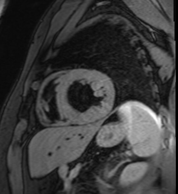
T1 fat sat short axis sequence used in heart imaging