MEDIC/MERGE/M-FFE MRI
Multiple Echo Data Image Combination (MEDIC) is a T2*-weighted spoiled gradient echo sequence. Multiple echoes acquired in one measurement are combined into an image. This sequence uses flow compensation unipolar frequency encoding gradients that remove CSF flow artifacts. MEDIC sequences produce a predominantly T2-weighted image.
Physics of the MEDIC MRI Sequence
The MEDIC MRI sequence relies on the principles of gradient echo (GRE) imaging and takes advantage of the phenomenon of signal decay over time, known as T2* relaxation. Here’s how it works:
Echo Train: The MEDIC sequence acquires multiple echoes during a single repetition time (TR). After the initial radiofrequency (RF) excitation pulse, a series of gradient echoes is collected with progressively increasing echo times (TE). This echo train allows for the acquisition of data at multiple echo times, each sensitive to different tissue properties.
T2 Sensitivity:* The varying echo times result in differences in T2* contrast within the acquired images. Tissues with different T2* values exhibit varying signal intensities in the final images. This property enables MEDIC to highlight specific tissue characteristics, such as fat-water differentiation or the detection of lesions with unique T2* properties.
Image Combination: After data acquisition, the multiple echoes are mathematically combined to create composite images with improved signal quality and contrast. This combination process helps reduce artifacts and enhance image fidelity.
MRI Image Appearance of MEDIC Sequence
The most straightforward way to recognize MEDIC images is by examining fluid-filled areas within the body, such as cerebrospinal fluid in the brain ventricles and spinal canal, synovial fluid in joints, or any abnormal fluid collections. In MEDIC images, fluids usually display a bright appearance, while muscles and fat typically manifest as various shades of gray.
Applications of the MEDIC MRI Sequence
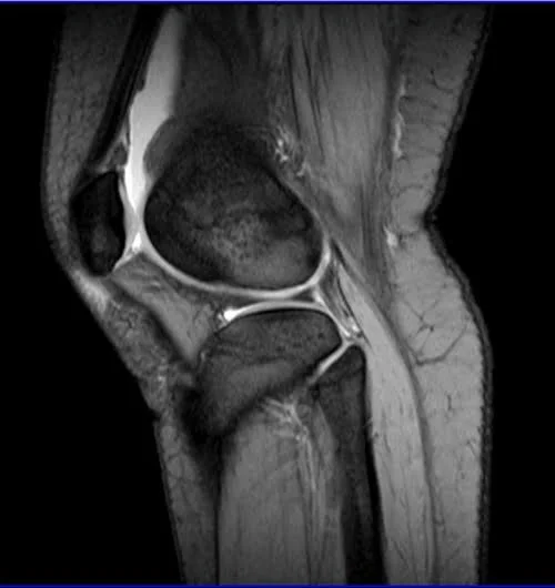
Cartilage Imaging: The high resolution and contrast make MEDIC particularly useful for visualizing hyaline cartilage, crucial for diagnosing osteoarthritis and other cartilage-related pathologies.
Musculoskeletal Imaging: MEDIC can beautifully depict musculoskeletal anatomy, identifying pathologies like tendon tears, ligament ruptures, and more.
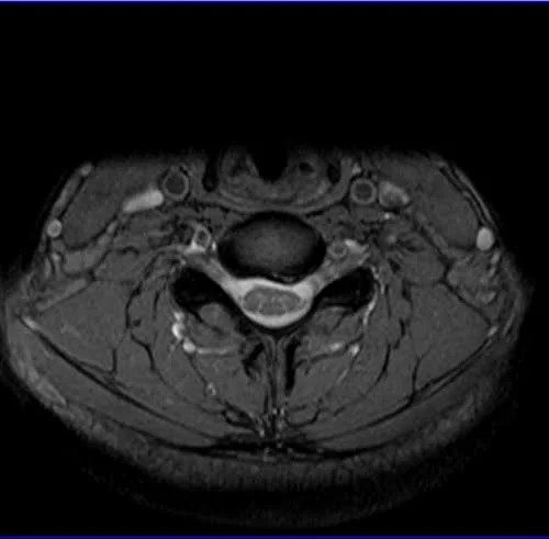
Cervical Spine Imaging: For cervical spine imaging, MEDIC sequences offer superior visualization of spinal cord structures, intervertebral discs, and surrounding tissues. They are instrumental in diagnosing spinal cord compression, herniated discs, and assessing spinal cord lesions or injuries.
Tissues and their MEDIC MRI appearance
Bone marrow – dark
Muscles – bright gray
Fat – gray (darker than muscle)
Moving blood– bright
Spinal cord – gray
Cartilage – bright
Fluids – bright
Bone – dark
Air – dark
Use
- Very useful for cervical spine imaging
- Very useful for cartilage imaging
- Useful for musculoskeletal imaging
- Very useful joints imaging
Axial MEDIC sequence used in cervical spine imaging
AXIAL MEDIC SEQUENCE USED IN WRIST IMAGING

SAGITTAL MEDIC SEQUENCE USED IN KNEE IMAGING
AXIAL MEDIC SEQUENCE USED IN ELBOW IMAGING

CORONAL MEDIC SEQUENCE USED IN SHOULDER
