Proton Density(PD) Fat Saturated
Proton Density (PD) fat-saturated MRI sequence is a specialized imaging sequence designed to improve contrast and visualization of non-fatty soft tissue structures. By selectively suppressing fat signals, this sequence enhances the visibility of edema, injuries, and pathologies in ligaments, bones, cartilage, and muscles. PD fat-saturated sequences are particularly advantageous in musculoskeletal imaging where fat can often overshadow subtle findings.
MRI Image Appearance of Proton Density(PD) Fat Saturated Image
The easiest way to identify proton density (PD) fat-saturated images is to look for adipose tissues in the body (e.g. subcutaneous fat and fat in bone marrow). Areas containing adipose tissues appear as dark on PD fat-saturated images. All other characteristics of the PD fat-saturated images remain the same as the PD image.
Tissues and their PD fat saturated images appearance
Musculoskeletal:
- Muscle: Intermediate
- Bone: Dark (low signal)
- Bone Marrow: Intermediate to dark
- Blood Vessels: Mostly Dark; Depending on flow characteristics, can be bright or dark
- Fat: Dark
- Ligaments: Intermediate to dark
- Nerve Roots: Intermediate
- Cartilage: Intermediate to dark
- Synovial Fluid: Bright
- Tendons: Intermediate to dark
Use
- Very useful for extremity imaging (e.g. ankle, knee, elbow shoulder and hips)
- Can be useful in thighs, lower legs, upper arms and forearms imaging
Pathology Appearance on Proton Density(PD) Fat Saturated Image
Proton Density (PD) weighted MRI sequences provide excellent visualization of soft tissue structures, particularly in musculoskeletal imaging. When combined with fat saturation, the contrast between certain tissues is further enhanced. Here’s a brief overview of how various pathologies might appear on PD fat-saturated scans:
Meniscal and Cartilage Lesions: In the context of knee imaging, meniscal tears and cartilage lesions can be detected as areas of increased signal on PD fat-saturated images. The fat saturation helps to suppress surrounding fatty marrow signals, making the tears more conspicuous.
Ligamentous and Tendon Injuries: Ligament and tendon tears or strains can be identified as areas of increased signal intensity increased signal intensity on PD fat-saturated images.
Ganglion Cysts and Bursitis: Fluid-filled structures such as ganglion cysts and bursal effusions will appear bright on PD fat-saturated images due to the high proton density of the fluid.
Bone Marrow Edema and Contusions: Areas of bone marrow edema, commonly seen after trauma or in inflammatory conditions, will appear as regions of increased signal intensity on PD fat-saturated images.
Synovitis: Inflamed synovium, which can contain both increased fluid and inflammatory cells, will be bright on PD fat-saturated images.
Muscle Injuries: Strains or tears in muscles can be identified as areas of increased signal.
Tumors: Soft tissue tumors can vary in appearance on PD fat-saturated images, depending on their content. Highly cellular or vascular tumors might show increased signal, while those with high fibrous content might appear darker.
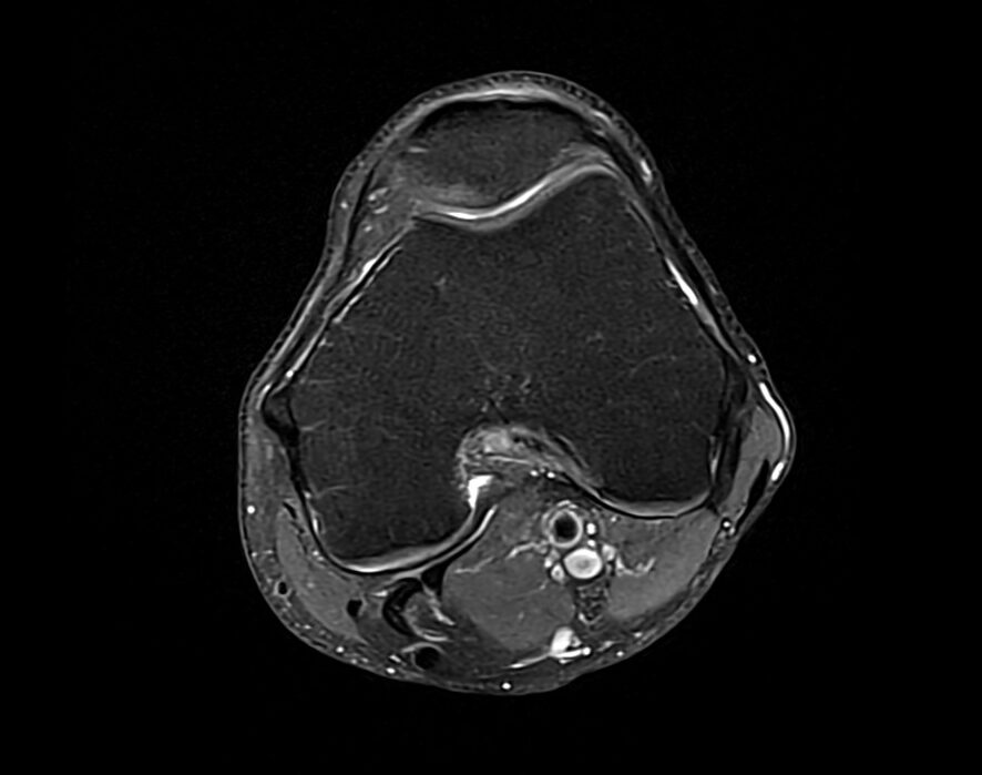
Proton Density(PD) Fat Saturated Axial Image of Knee
Proton Density(PD) Fat Saturated Coronal Image of Knee

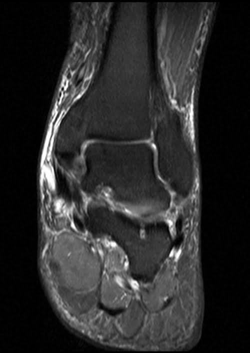
Proton Density(PD) Fat Saturated Coronal Image of Ankle