Phase-contrast (PC) flow MRI: Physics and Applications
Phase-contrast (PC) flow MRI is a specific type of magnetic resonance imaging (MRI) technique that is used to visualize and quantify dynamic fluid flow within the body. This technique is particularly useful because it allows for the non-invasive measurement of the velocity of moving fluids, such as blood or cerebrospinal fluid (CSF).
Physics of Phase-Contrast Flow MRI
The physics behind phase-contrast flow MRI involves the phenomenon of phase shift. When protons within flowing blood encounter a magnetic field gradient, they experience a phase shift proportional to their velocity. By applying bipolar gradient pulses perpendicular to the direction of flow, phase shifts are induced in moving protons. These phase shifts manifest as differences in the phase of the MRI signal, which can be detected and used to calculate the velocity of blood flow.
Here’s a breakdown of the physics involved:
Gradient Magnetic Fields: In addition to the main magnetic field, PC-MRI employs gradient magnetic fields that are temporally and spatially varied. These gradients are applied in specific directions (usually aligned with the expected direction of flow).
Phase Shifts: When these gradient fields are applied, moving protons acquire a phase shift proportional to their velocity. This phase shift occurs because the moving protons experience a different magnetic environment along their path, compared to stationary protons.
Velocity Encoding (VENC): The velocity of moving fluid is encoded into the MRI signal by adjusting the gradients. The maximum velocity that can be accurately measured without aliasing (where higher velocities might incorrectly appear as lower velocities) is determined by a parameter known as VENC. Choosing an appropriate VENC is crucial for accurate velocity measurements; if it’s too low, velocity aliasing occurs, and if it’s too high, the sensitivity to slow flow decreases.
Image Appearance
In phase-contrast images, the intensity of each pixel corresponds to the velocity and direction of flow relative to the orientation of the applied gradient. The images are typically presented as:
Re-phased Image (Magnitude of Flow Compensated Signal):
This image captures the magnitude of the flow-compensated signal, where flow velocities are adjusted to enhance visibility against the static background tissues. It highlights high signal intensity for flow areas while keeping the background visible, useful for evaluating overall flow dynamics. Here’s a simple breakdown of the image appearance:
- Flow Signal: High signal intensity.
- Background Visibility: The background structures remain visible in this type of image. The purpose of the re-phased image is to measure the signal where flow has been compensated. This means that the flow velocities are adjusted to appear as high signal areas, facilitating the identification of flow dynamics against the static tissues.
Magnitude Image (Magnitude of Difference Signal):
The magnitude image is derived from the magnitude of the difference between phase-shifted signals. It features high signal intensity for flow, irrespective of the direction, with a suppressed background. This enhancement focuses on clearly delineating flow from the surrounding tissues, emphasizing the areas of fluid movement. Here’s a simple breakdown of the image appearance:
- Flow Signal: High signal intensity regardless of the direction of flow.
- Background Suppression: In this image, the background is intentionally suppressed to enhance the visibility of the flow. The magnitude image is created by calculating the absolute difference between the phase-shifted and flow-compensated images. This process accentuates areas of flow, making them distinctly brighter than non-moving tissues, thus focusing the analysis strictly on fluid dynamics.
Phase Image (Phase of Difference Signal):
In the phase image, the signal intensity varies with the direction of flow—forward flow yields a high signal and reverse flow a low signal, against a mid-grey background. This directional sensitivity is critical for assessing the velocity and directionality of fluid movements within the body, aiding in precise diagnostics. Here’s a simple breakdown of the image appearance:
- Flow Signal: Dependent on the direction of the flow; forward flow is depicted with high signal, whereas reverse flow appears as low signal.
- Background: Typically displayed in mid-grey, which helps to differentiate the flow direction against a neutral backdrop. The phase image encodes the velocity of moving fluids by varying the signal intensity according to the direction and speed of flow. This directional sensitivity is crucial for detailed assessments of fluid dynamics, such as determining the direction of blood flow in cardiovascular assessments.
Re-phased Image
Magnitude Image
Phase Image
Uses of Phase-Contrast MRI
Phase-Contrast Flow MRI (PC-MRI) is a versatile imaging technique used in various medical and research fields due to its ability to non-invasively measure and visualize fluid flow within the body. Here are some key applications of this technology:
Cardiovascular Applications
PC-MRI is extensively used in cardiology for assessing blood flow dynamics within the heart and major vessels:
- Valvular Heart Disease: It helps in quantifying the severity of valvular regurgitation and stenosis by measuring the flow across the affected valves.
- Congenital Heart Defects: PC-MRI is crucial for the evaluation of congenital heart diseases, such as septal defects and anomalies in pulmonary or systemic circulation.
- Vascular Studies: It assesses the hemodynamics of vascular diseases, including aneurysms and dissections, helping to understand the risk and monitor treatment outcomes.
- Pulmonary Vascular and Airway Diseases: It can assess blood flow in pulmonary arteries and veins, useful in diseases like pulmonary hypertension.
Phase-contrast (PC) sequence used in in-plane aortic flow imaging
Re-phased Image

Magnitude Image

Phase Image
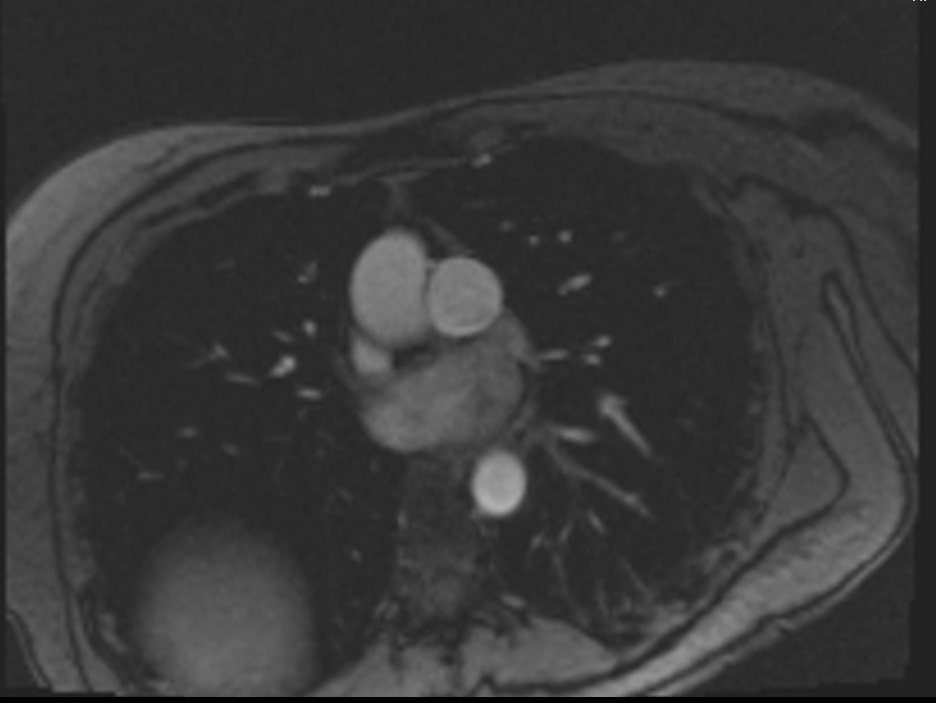
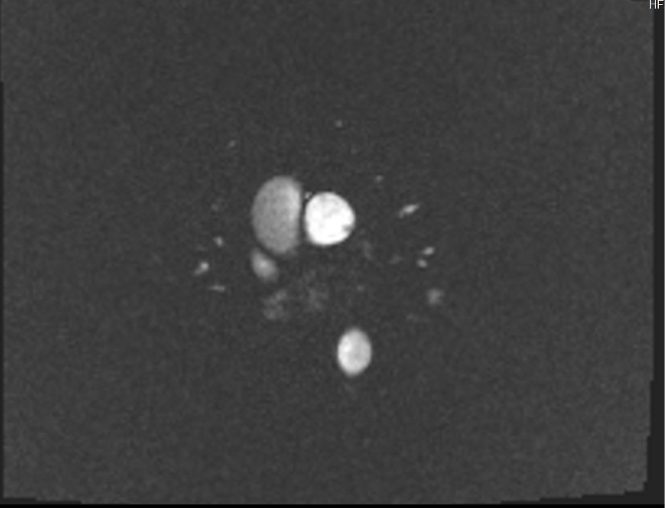
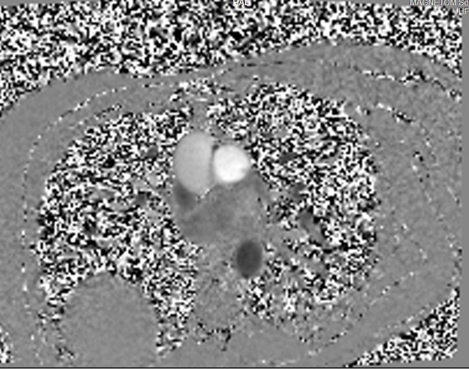
Phase-contrast (PC) sequence used in through-plane pulmonary artery imaging
Re-phased Image
Magnitude Image
Phase Image
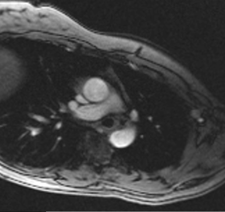
Phase-contrast (PC) sequence used in through-plane aortic artery imaging
Re-phased Image
Magnitude Image

Phase Image

Neurological Applications
In neurology, PC-MRI is used to study the flow of cerebrospinal fluid (CSF) and assess vascular conditions:
- CSF Flow Analysis: PC-MRI measures the dynamics of CSF flow in conditions like hydrocephalus, Chiari malformation, and spinal cord cysts, aiding in their diagnosis and treatment planning.
- Arteriovenous Malformations (AVMs): It can be used to visualize and quantify the flow within AVMs, which is crucial for pre-surgical planning and assessing the efficacy of treatment.
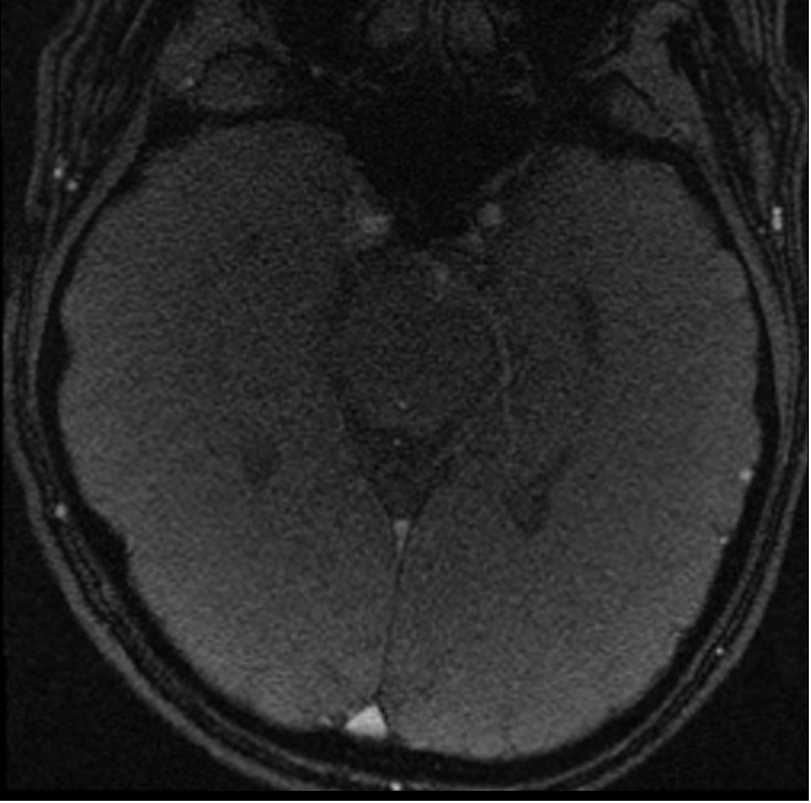
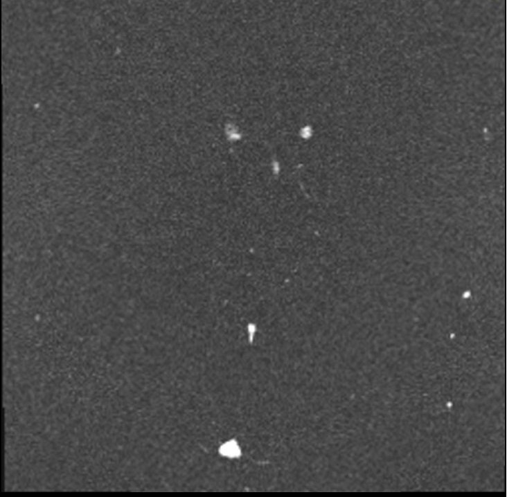
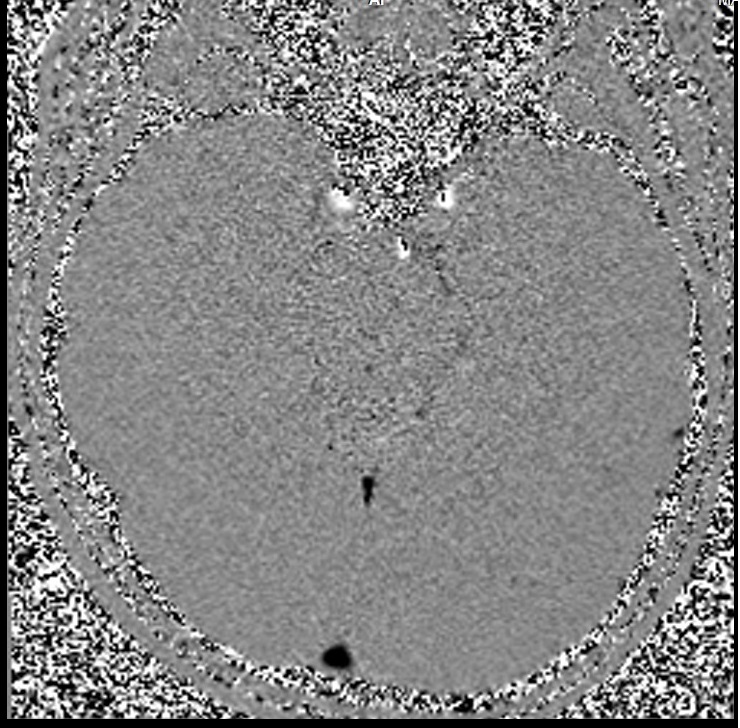
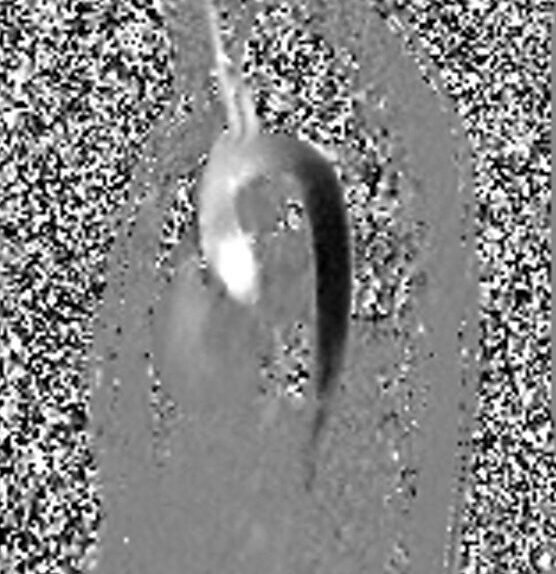
Phase-contrast (PC) sequence used in CSF flow imaging
Re-phased Image
Magnitude Image
Phase Image
Research and Functional Studies
- Hemodynamic Research: Researchers use PC-MRI to study the hemodynamics within vascular diseases, exploring how changes in blood flow dynamics contribute to conditions like arterial stenosis or vascular aneurysms.
- Muscle and Joint Kinetics: PC-MRI can measure the velocities of tissues during movement, providing insights into muscle contractions and joint mechanics. This is particularly useful in sports science and rehabilitation medicine.
- Kidney and Liver Blood Flow: PC-MRI is used to assess blood flow in renal and hepatic arteries and veins. This information is important in conditions like renal artery stenosis or cirrhosis, where blood flow is significantly affected.
- 4D Flow MRI: This extension of PC-MRI captures three-dimensional flow dynamics over time, providing detailed visualization and analysis of complex blood flow patterns, particularly useful in comprehensive vascular assessments and surgical planning.
- Perfusion Studies: It assists in calculating perfusion in tissues by measuring the arterial input function, which is essential for dynamic contrast-enhanced studies in organs like the brain, heart, and tumors.
References
- Battal, B., Kocaoglu, M., Bulakbasi, N., Husmen, G., Sanal, H. T., & Tayfun, C. (2011). Cerebrospinal fluid flow imaging by using phase-contrast MR technique. The British Journal of Radiology, 84(1004), 758–765. https://doi.org/10.1259/bjr/66206791
- Wymer, D. T., Patel, K. P., Burke III, W. F., & Bhatia, V. K. (2020). Phase-Contrast MRI: Physics, Techniques, and Clinical Applications. RadioGraphics, 40(1). https://doi.org/10.1148/rg.2020190039
- Nayak, K. S., Nielsen, J.-F., Bernstein, M. A., Markl, M., Gatehouse, P. D., Botnar, R. M., Saloner, D., Lorenz, C., Wen, H., Hu, B. S., Epstein, F. H., Oshinski, J. N., & Raman, S. V. (2015). Cardiovascular magnetic resonance phase contrast imaging. Journal of Cardiovascular Magnetic Resonance, 17(1), 71. https://doi.org/10.1186/s12968-015-0172-5