T1 SE / T1 TSE / T1 FSE Fat Saturated Post Contrast
A T1 fat-saturated post contrast sequence is a specific magnetic resonance imaging (MRI) sequence utilized to improve the visibility of certain anatomical structures and pathological changes within the body. This sequence incorporates three essential components:
T1-weighted imaging (T1W): Refers to the time taken for tissue to recover 63% of its longitudinal magnetization after being disturbed by an external magnetic pulse. T1-weighted images highlight differences in the longitudinal relaxation times of tissues, producing images where fat appears bright.
Fat Saturation (FAT SAT): A technique applied to suppress the signal from fat tissues. This makes it easier to detect and differentiate lesions, especially when they are located near fatty structures.
Post Contrast: Involves the administration of a contrast agent, usually gadolinium-based, which helps to enhance the visibility of vascular structures, tumors, and areas of inflammation.
MRI image appearance
The easiest way to identify T1 weighted fat saturated post gadolinium images is to look for adipose tissues (e.g. subcutaneous fat and fat in bone marrow) and blood vessels in the body (e.g arteries and veins in the brain, neck, chest, abdomen, upper limbs and lower limbs). Areas contain adipose tissues appear dark on T1 weighted fat saturated post gadolinium images. Blood vessels and pathologies with high vascularity appear bright on T1 weighted fat saturated post gadolinium images.
Tissues and their T1 fat saturated post gadolinium appearance
Brain:
- CSF: Dark.
- White Matter: Intermediate.
- Gray Matter: Intermediate to dark.
- Bone (skull): Dark.
- Bone Marrow: Intermediate..
- Blood Vessels: Mostly Bright. Depending on flow characteristics, can be Dark.
- Pituitary Gland: Bright.
- Choroid Plexus: Bright.
- Cerebellum: Gray matter Darker than white matter.
- Brain Stem: Intermediate
- Sinuses: Dark (air-filled).
- Thalamus, Putamen, Hippocampus, Caudate Nucleus: Intermediate
- Corpus Callosum: Intermediate
- Pineal Gland: Intermediate, may show enhancement depending on pathology.
Spine:
- Spinal Cord: Intermediate to bright.
- CSF: Dark.
- Bone: Dark.
- Bone Marrow: Intermediate.
- Intervertebral Disc: Nucleus pulposus and annulus are Intermediate tobright.
- Ligaments: Intermediate to Dark.
- Nerve Roots: Intermediate.
Abdomen and Pelvis:
- Liver: Intermediate to bright.
- Gallbladder and Common Bile Duct: Dark
- Spleen: Bright.
- Kidney: Bright.
- Ureters: Intermediate to bright.
- Pancreas: Bright.
- Urinary Bladder: Bright (when filled with urine).
- Prostate: Bright.
- Uterus: Bright.
Musculoskeletal:
- Muscle: Intermediate to dark
- Bone: Dark (low signal)
- Bone Marrow: Intermediate
- Blood Vessels: Mostly bright
- Fat: Dark
- Ligaments: Intermediate to dark
- Nerve Roots: Intermediate
- Cartilage: Intermediate
- Synovial Fluid: Dark
- Tendons: Intermediate to dark
Use
- Very useful for pituitary imaging
- Very useful for spine imaging
- Very useful for pelvic imaging
- Very useful for anterior neck, orbits and face imaging
- Very useful for any musculoskeletal imaging
- Very useful for extremity imaging
Pathological appearance
In a T1 fat-saturated post-gadolinium MRI sequence, various pathologies demonstrate unique appearances based on their inherent tissue characteristics and vascularity:
Adipose Tissue Content: Pathologies that predominantly consist of fat, such as lipomas, will appear dark. The fat saturation technique suppresses the signal from fat, making these lesions contrast sharply against other tissues.
Hypervascular Pathologies: Lesions that have a high blood supply, or hypervascularization, will show bright enhancement after gadolinium administration. This includes tumors like hemangioma, lymphangioma, hemangioendothelioma, Kaposi sarcoma, angiosarcoma, and hemangioblastoma. Inflammatory processes like discitis, meningitis, synovitis, arthritis, and osteomyelitis also demonstrate pronounced enhancement due to increased blood flow.
Non-vascular Pathologies: Pathological processes that lack significant blood vessels will not take up the gadolinium contrast and, hence, will remain dark on a T1 fat-saturated post-gadolinium image.
T1 fat-saturated coronal post-contrast sequence used in pituitary gland imaging

T1 fat-saturated axial post contrast sequence used in orbits imaging

T1 fat-saturated axial post contrast sequence used in neck imaging.

T1 fat-saturated sagittal post-contrast sequence used in cervical spine imaging

T1 fat-saturated sagittal post-contrast sequence used in lumbar spine imaging

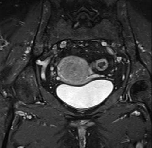
T1 fat-saturated axial (uterus) post-contrast sequence used in female pelvic imaging
T1 fat-saturated axial post-contrast sequence used in female urethra imaging.

Post-contrast fat-saturated axial T1-weighted sequence used in hip arthrography imaging.

Post-contrast fat-saturated axial T1-weighted sequence used in shoulder arthrography imaging.
