MRI Flow Artifacts
Flow artifacts in MRI refer to distortions or signal changes in the images caused by the movement of fluids, such as cerebrospinal fluid (CSF) and blood flow. These artifacts can result in blurring, ghosting, or signal loss in the images, making it challenging to accurately interpret the anatomical structures.

Blood Flow Related Artifacts
Blood flow artifacts in MRI refer to image distortions or signal loss caused by the motion of blood within vessels. Blood flow artifacts is often seen in regions where there is a pulsatile blood flow such as the heart, large vessels, and sometimes the brain. Blood flow can create several types of artifacts in MRI images. The most common types of artifacts created by blood flow include:
Flow Void: In spin-echo sequences (but not GRE) fast flowing blood may appear dark or black: as blood moves, some spins may enter or leave the selected slice in the time between administration of the 90° pulse and the 180° pulse. As the spins only receive one of these pulses, either excitation or refocusing does not occur and a spin echo signal cannot be produced.

Slower flowing blood will appear brighter in spin echo sequences as more spins will be present in the slice for both 90° and 180° pulses and will be able to produce a signal. The differential appearance of slow and fast flowing blood in spin echo may aid diagnosis of thrombosis, aneurysm or vascular malformation.

To avoid the flow void artifact, several techniques can be used
Thicker slices can be used, which will increase the likelihood of spins being present in the slice for both the 90° and 180° pulses, allowing them to produce a signal and reduce the appearance of flow void.
Lower TE can also be used, which will reduce the time between the 90° and 180° pulses and decrease the likelihood of spins moving in and out of the slice, thereby reducing the occurrence of flow void.
Additionally, gradient moment nulling techniques can be used to compensate for the difference in phase between static and flowing spins in a slice, which can help to reduce the occurrence of flow void.
NEX (Number of Excitations): NEX refers to the number of times the MRI scan is repeated to acquire multiple signal averages. Increasing the NEX can help improve the signal-to-noise ratio (SNR) by reducing random noise. This, in turn, can enhance image quality and reduce flow artifacts.
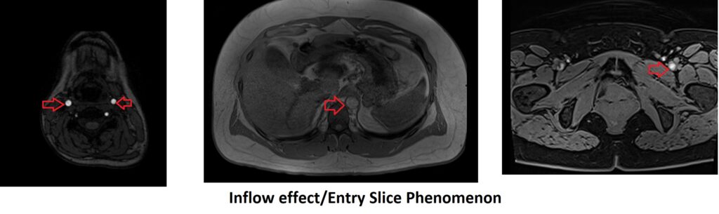
Inflow effect/Entry Slice Phenomenon: This effect creates a bright signal, especially in GRE imaging and in entry slices of spin echo sequences. Fresh, unsaturated spins in blood flow into a slice. Stationary spins in the same slice which have not completed T1 relaxation between RF pulses remain partially saturated and will emit less signal after the next RF pulse than the relaxed spins in flowing blood. Blood therefore looks comparatively bright. This effect is the basis of Time-of-flight (TOF) MR angiography.
To avoid the Entry Slice Phenomenon artifact, several techniques can be used:
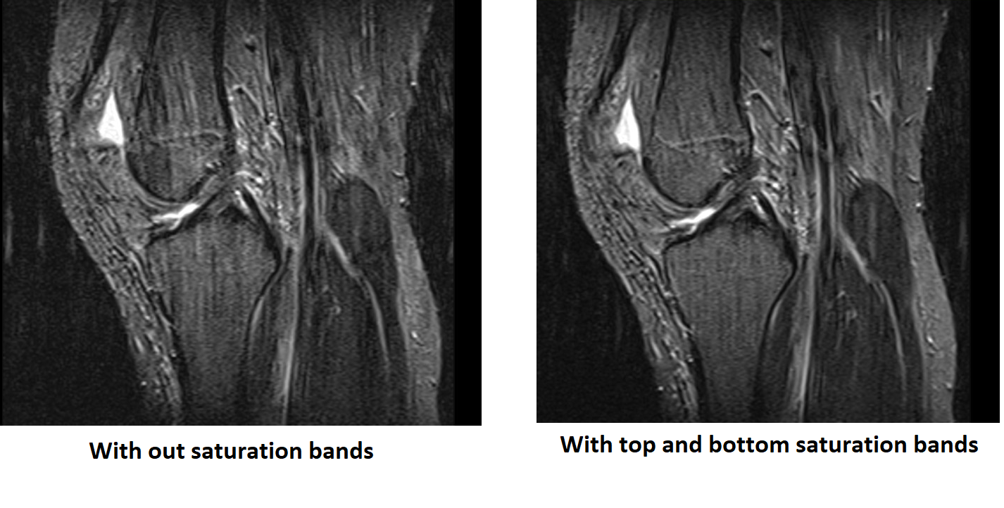
Saturation bands: Saturation bands are applied outside the region of interest (ROI) to suppress the signal from flowing blood. They are typically placed upstream and downstream of the ROI, effectively “tagging” the flowing blood. This reduces the contribution of flowing blood to the resulting image and minimizes flow artifacts.
NEX (Number of Excitations): NEX refers to the number of times the MRI scan is repeated to acquire multiple signal averages. Increasing the NEX can help improve the signal-to-noise ratio (SNR) by reducing random noise. This, in turn, can enhance image quality and reduce flow artifacts.
Use selective saturation pulses: One way to reduce the inflow effect is by using selective saturation pulses or pre-pulses to saturate the flowing blood before the imaging pulse sequence begins. This can help to eliminate the unsaturated spins in the blood, reducing the inflow effect.
Use gradient-echo sequence: Another technique to minimize the inflow effect is to use a gradient-echo sequence instead of a spin-echo sequence. In gradient-echo imaging, the magnetization of the flowing blood is not saturated as much as in spin-echo imaging, which reduces the inflow effect.
Use gradient-echo sequence: Flow compensation techniques, such as gradient moment nulling, can also help to reduce the inflow effect. These techniques use additional radiofrequency pulses to compensate for the difference in phase between static and flowing spins in a slice, helping to reduce artifacts caused by flowing blood.
Adjusting the imaging parameters : Adjusting the imaging parameters, such as flip angle, repetition time (TR), and echo time (TE), can also help to minimize the inflow effect. For example, a longer TR can reduce the inflow effect by allowing the magnetization to recover before the next pulse sequence begins. Similarly, a shorter TE can reduce the inflow effect by minimizing the time between the RF pulse and the echo, which can reduce the number of unsaturated spins in the flowing blood.
Displacement artifact: Flow Displacement artifact in MRI occurs when there is a time difference between the phase-encoding (PE) and frequency-encoding (FE) steps during image acquisition. This time difference can cause blood to move between the PE and FE steps, leading to misregistration of the true position of the blood vessel on the image and the appearance of the blood outside the vessel.

There are several ways to minimize the flow displacement artifact in MRI:
Saturation bands: Saturation bands are applied outside the region of interest (ROI) to suppress the signal from flowing blood. They are typically placed upstream and downstream of the ROI, effectively “tagging” the flowing blood. This reduces the contribution of flowing blood to the resulting image and minimizes flow artifacts.
NEX (Number of Excitations): NEX refers to the number of times the MRI scan is repeated to acquire multiple signal averages. Increasing the NEX can help improve the signal-to-noise ratio (SNR) by reducing random noise. This, in turn, can enhance image quality and reduce flow artifacts.
Phase direction: The phase direction in which the MRI acquisition is performed can influence the appearance and severity of flow artifacts. By altering the direction of flow relative to the imaging plane, it is possible to minimize the impact of these artifacts. In the case of knee imaging in the sagittal plane, changing the phase direction from anterior-posterior to head-to-feet can help reduce flow-related artifacts.

Reduce the TE (echo time): Shortening the TE reduces the time between the PE and FE steps, minimizing the time for flow effects to occur.
Increase the voxel size: Increasing the voxel size reduces the time required for image acquisition, reducing the time for flow effects to occur.
Use flow compensation techniques: Flow compensation techniques use additional gradient pulses to compensate for the differences in phase between static and moving spins, which can minimize the flow displacement artifact.
Use parallel imaging techniques: Parallel imaging techniques use multiple receiver coils to acquire data simultaneously, which can reduce the time required for image acquisition and minimize the flow displacement artifact.
Aliasing artifact: This artifact occurs when the velocity of blood flow exceeds the maximum velocity encoding limit of the imaging sequence. This causes the MRI signal to be wrapped around, resulting in a false appearance of blood vessels.

There are several ways to minimize the flow Aliasing artifact in MRI:
There are several ways to minimize the flow Aliasing Artifact in MRI
Saturation bands: Saturation bands are applied outside the region of interest (ROI) to suppress the signal from flowing blood. They are typically placed upstream and downstream of the ROI, effectively “tagging” the flowing blood. This reduces the contribution of flowing blood to the resulting image and minimizes flow artifacts.
NEX (Number of Excitations): NEX refers to the number of times the MRI scan is repeated to acquire multiple signal averages. Increasing the NEX can help improve the signal-to-noise ratio (SNR) by reducing random noise. This, in turn, can enhance image quality and reduce flow artifacts.
Oversampling: Oversampling refers to acquiring more data points in the phase-encoding direction during the MRI scan. This helps to increase the spatial resolution and reduce the effect of flow artifacts. The increased number of data points allows for better separation of flowing blood from stationary tissues.
Flow-related aliasing artifact can be avoided or minimized by adjusting the imaging parameters such as increasing the velocity encoding (VENC) value, reducing the voxel size, changing the phase encoding direction or decreasing the repetition time (TR).
Velocity encoding (VENC) is the maximum velocity that can be measured without aliasing. By increasing the VENC value, higher velocities can be measured without aliasing, thus reducing the occurrence of the artifact. However, increasing the VENC value may also decrease the signal-to-noise ratio (SNR) and decrease the spatial resolution.
Reducing the voxel size can also help minimize the occurrence of flow-related aliasing artifact. By decreasing the voxel size, the amount of blood that enters the voxel is reduced, which decreases the likelihood of aliasing.
Decreasing the repetition time (TR) can also help reduce the occurrence of flow-related aliasing artifact. By decreasing the TR, there is less time for the blood to move between acquisitions, which reduces the likelihood of aliasing.
Flow-related aliasing artifact can be avoided by changing the phase encoding direction. By changing the direction of the phase encoding gradient, the direction of flow can also be changed, which can help to minimize the flow-related aliasing artifact. This is because the artifact occurs when the flow is perpendicular to the phase encoding direction, and changing the direction can help to align the flow more parallel to the phase encoding direction, resulting in less aliasing.
CSF Flow Related Artifacts
CSF flow artifact is caused by the movement of CSF between the time of excitation and signal acquisition, resulting in a phase shift in the MRI signal. This can be particularly problematic in certain MRI sequences, such as fast spin echo (FSE) and gradient echo (GRE) sequences, which are sensitive to phase shifts.
There are several types of artifacts that can be created by cerebrospinal fluid (CSF) flow during MRI imaging. Some common examples include:
Ghosting artifact: This is caused by CSF pulsation, which can create ghost images of brain structures. These ghost images can be particularly problematic in functional MRI studies, where they can be misinterpreted as neuronal activation.

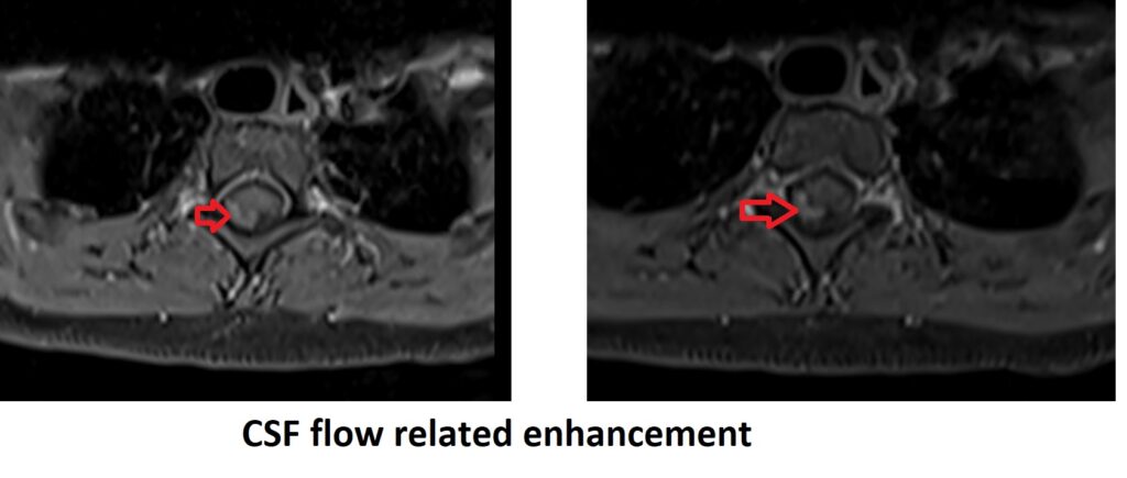
Flow-related enhancement: In post-contrast MRI studies, the flow of CSF can sometimes create areas of enhancement that mimic pathology. This can be especially problematic in the evaluation of brain and spinal cord pathologies.
Flow void artifact: Similar to the flow void artifact created by blood flow, this artifact appears as signal loss in areas where CSF is flowing quickly, such as the cerebral aqueduct.

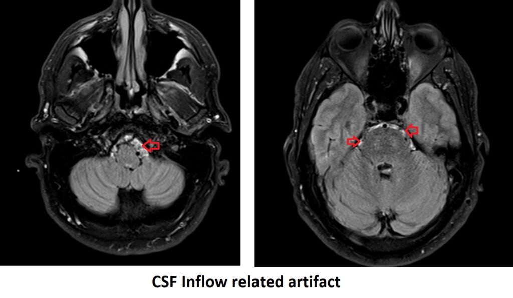
Inflow artifact: When CSF enters a region of the brain during a pulse sequence, it can create an artifact that appears as a bright signal. This can be useful in some MRI studies, but can also be a source of confusion if not recognized.
There are several ways to minimize or avoid CSF Flow Artifact in MRI
Use a higher receiver bandwidth: This can reduce the chemical shift artifact that can occur with CSF flow, as the wider bandwidth will allow the receiver to detect the signal from both the fat and water protons.
Using flow suppression techniques: These techniques involve the use of additional RF pulses or gradients to suppress the signal from the flowing CSF and improve image quality. For example, the double inversion recovery (DIR) sequence can suppress CSF signal, and the motion-sensitized driven-equilibrium (MSDE) sequence can suppress signal from moving fluids.
Using phase encoding direction: Changing the phase encoding direction can help to avoid CSF flow artifact. In some cases, using a 180-degree phase shift can help to suppress the artifact.
Use a longer echo time (TE): A longer TE can also reduce the chemical shift artifact by allowing more time for the phase differences between fat and water to be resolved.
Use flow compensation techniques: Flow compensation techniques, such as bipolar gradients or gradient moment nulling, can be used to compensate for the effects of CSF flow, reducing the artifact.
Change the imaging plane orientation: Changing the imaging plane orientation can sometimes help to avoid CSF flow artifact. For example, imaging in the axial plane instead of the sagittal or coronal planes can reduce the effects of CSF flow.
Use of motion correction techniques: Motion correction techniques such as Radial k space sampling ,navigator echoes and prospective motion correction can be used to reduce or eliminate CSF flow artifacts..
Saturation bands: Placing saturation bands around the area of interest can help to suppress CSF flow signals. The saturation bands act like a filter, preventing the flow signal from reaching the region of interest.
References
- Song AW, Huang W, Van Gelderen P. Phase-cycled balanced steady-state free precession for arterial spin labeling MRI. Magn Reson Med. 2011;66(2):405-410. doi:10.1002/mrm.22824
- Haacke EM, Xu Y, Cheng YCN, et al. Susceptibility weighted imaging (SWI). Magn Reson Med. 2004;52(3):612-618. doi:10.1002/mrm.20198
- Krupa P, Pattany PM, Ciancibello L, et al. Cerebrospinal fluid flow imaging by means of MRI using a multiphase-balanced steady-state free precession sequence. AJR Am J Roentgenol. 2003;180(3):745-748. doi:10.2214/ajr.180.3.1800745
- Haacke EM, Brown RW, Thompson MR, Venkatesan R. Magnetic Resonance Imaging: Physical Principles and Sequence Design. 2nd ed. Wiley-Liss; 1999.
- Bammer R. Basic principles of diffusion-weighted imaging. Eur J Radiol. 2003;45(3):169-184. doi:10.1016/s0720-048x(02)00303-6
- Andre JB, Bammer R. Advanced MR diffusion imaging. Top Magn Reson Imaging. 2008;19(2):97-114. doi:10.1097/rmr.0b013e318180c2fd
- Schaefer PW, Grant PE, Gonzalez RG. Diffusion-weighted MR imaging of the brain. Radiology. 2000;217(2):331-345. doi:10.1148/radiology.217.2.r00nv08331
- Watanabe T, Frahm J, Michaelis T. Dynamic imaging of perfusion using a split-echo cine imaging technique. Magn Reson Med. 2001;46(3):522-529. doi:10.1002/mrm.1209
- Johnson KM, Fain SB, Schiebler ML, Nagle SK. Optimized 3D ultrashort echo time pulmonary MRI. Magn Reson Med. 2013;69(4):1231-1237. doi:10.1002/mrm.24375
- Wu M, Chang WC, Yang SC, et al. Phase-sensitive inversion recovery for flow artifact reduction in intracranial 3D TOF MR angiography: initial experience. AJNR Am J Neuroradiol. 2008;29(8):1524-1528. doi:10.3174/ajnr.A1141
- Zwart NR, Pipe JG. Flow compensation in MRI. J Magn Reson Imaging. 2015;42(3):586-598.
- Vymazal J. Clinical applications of the phase contrast techniques in MR imaging. Eur Radiol. 1999;9(7):1292-1306.
- Farzaneh F, Khodarahmi I, Akhlaghpoor S, Mohajer K. Artifacts in MRI: A review. Asian J Med Radiol. 2014;2(2):41-49.
- Reeder SB, Pineda AR, Wen Z, et al. Iterative decomposition of water and fat with echo asymmetry and least-squares estimation (IDEAL): application with fast spin-echo imaging. Magn Reson Med. 2005;54(3):636-644.
- Lauenstein TC, Sharma P, Hughes T, Heberlein K, Tudorascu D, Martin DR. Evaluation of optimized inversion-recovery fat-suppression techniques for T2-weighted abdominal MR imaging. J Magn Reson Imaging. 2008;28(2):421-426.
- Bydder GM, Hamilton G, Yokoo T, et al. Relaxation effects in the quantification of fat using gradient echo imaging. Magn Reson Imaging. 2008;26(3):347-359.
- Nayak KS, Cunningham CH, Santos JM, Pauly JM. Real-time cardiac MRI at 3 Tesla. Magn Reson Med. 2004;51(4):655-660.
- Goksel O, Buechler P, Reyes M. Phase residual errors in quantitative flow imaging with phase-contrast MRI. Magn Reson Med. 2012;67(2):510-517.