Anal Fitsula MRI Protocol and Planning
Indications for fistula MRI scans
- For the assessment of the activity of perianal fistulas in the course of Crohn's disease
- For pre-operative evaluation of perineal abscess
- For pre-operative evaluation of perineal fistula
- To check the anatomy of anal region
- To check the extend of fistulas
- Causes of perianal fistulas
Contraindications
- Any electrically, magnetically or mechanically activated implant (e.g. cardiac pacemaker, insulin pump biostimulator, neurostimulator, cochlear implant, and hearing aids)
- Intracranial aneurysm clips (unless made of titanium)
- Pregnancy (risk vs benefit ratio to be assessed)
- Ferromagnetic surgical clips or staples
- Metallic foreign body in the eye
- Metal shrapnel or bullet
Patient preparation for fistula MRI scans
- A satisfactory written consent form must be taken from the patient before entering the scanner room
- Ask the patient to remove all metal objects including keys, coins, wallet, cards with magnetic strips, jewellery, hearing aid and hairpins
- Ask the patient to undress and change into a hospital gown
- If possible provide a chaperone for claustrophobic patients (e.g. relative or staff )
- Offer earplugs or headphones, possibly with music for extra comfort
- Explain the procedure to the patient
- Instruct the patient to keep still
- Note the hight and weight of the patient
Please check our new video tutorial for protocols and planning
Positioning for fistula MRI scans
- Position the patient in supine position with head pointing towards the magnet (head first supine)
- Position the patient over the spine coil and place the body coil over the pelvis ( iliac crest down to mid thigh)
- Securely tighten the body coil using straps to prevent respiratory artefacts
- Give a pillow under the head for extra comfort
- Centre the laser beam localiser over symphysis pubis (4 inches below iliac crest)
- Avoid placing cushions under the legs as it may elevate the bottom of the pelvis away from the coil.

Recommended MRI Fistula Protocols and Planning
localiser
To localize and plan the sequences, it is essential to initially acquire a three-plane T2 HASTE localizer. These fast single-shot localizers have an acquisition time of under 25 seconds and are highly effective in accurately localizing pelvic structures.

localiser 2
For proper planning of fistula scans, it is necessary to include a second two-plane localizer. The two-plane localizer should be positioned on the sagittal plane. Align the planning block parallel to the anal canal for the coronal plane and perpendicular to the anal canal for the axial plane.

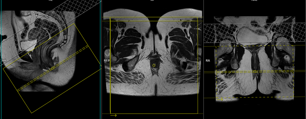
T2 tse DIXON sagittal 3mm
Plan the sagittal slices on the coronal plane and angle the planning block parallel to the anal canal (i.e., parallel to the pubic symphysis). Verify the planning block in the other two planes. Provide an appropriate angle in the axial plane, parallel to the line connecting the pubic symphysis and the anal canal. The slices should adequately cover the entire pelvis, spanning from the right ischial tuberosity to the left ischial tuberosity. Use a field of view (FOV) large enough to encompass the entire buttock region.

Parameters
TR 5000-6000 | TE 90-120 | SLICE 3 MM | FLIP 151 | PHASE H>F | MATRIX 320X320 | FOV 240-280 | GAP 10% | NEX(AVRAGE) 2 |
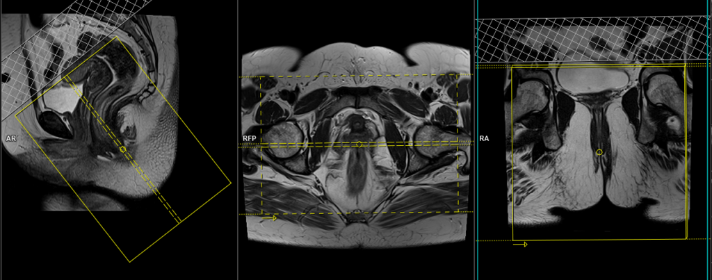
T2 tse DIXON\STIR axial oblique 3mm
Plan the axial slices on the sagittal plane; angle the planning block perpendicular to the anal canal. Verify the planning block in the other two planes. Ensure an appropriate angle is applied in the coronal plane, which should be perpendicular to the anal canal. The slices should adequately cover the entire buttock area from the middle of the rectum down to the skin level of the buttock. This comprehensive coverage is crucial as most fistulas extend and open at the skin surface of the buttock. To minimize ghosting artifacts caused by peristalsis and breathing, consider using a saturation band over the axial block.
Parameters
TR 5000-6000 | TE 110 | FLIP 150 | NEX 2 | SLICE 3 MM | MATRIX 256X256 | FOV 250-300 | PHASE R>L | GAP 10% | FAT SAT DIXON |
T2 tse DIXON\STIR coronal oblique 3mm
Plan the coronal slices on the sagittal plane; angle the planning block parallel to the anal canal. Check the planning block in the other two planes. An appropriate angle must be given in the axial plane (Parallel to the right and left hip or ischial tuberosity). Slices must be sufficient to cover the entire buttock, starting two slices in front of the symphysis pubis and extending up to the level of the sacrum. To minimize ghosting artifacts caused by peristalsis and breathing, consider using a saturation band over the coronal block.
Parameters
TR 5000-6000 | TE 110 | FLIP 150 | NEX 2 | SLICE 3 MM | MATRIX 256X256 | FOV 250-300 | PHASE R>L | GAP 10% | FAT SAT DIXON |
Optional Scans
T2 SPACE 3D axial oblique 1mm
Plan the axial slices on the sagittal plane; angle the planning block perpendicular to the anal canal. Verify the planning block in the other two planes. Ensure an appropriate angle is applied in the coronal plane, which should be perpendicular to the anal canal. The slices should adequately cover the entire buttock area from the middle of the rectum down to the skin level of the buttock. This comprehensive coverage is crucial as most fistulas extend and open at the skin surface of the buttock. To minimize ghosting artifacts caused by peristalsis and breathing, consider using a saturation band over the axial block.

Parameters
TR 1500-2000 | TE 97 | FLIP 170 | NEX 1.6 | SLICE 1 MM | MATRIX 256X256 | FOV 270-300 | PHASE A>P | IPAT GRAPPA2 | OVERSAMPLE 40% |