MRI Magnetic Susceptibility Artifact
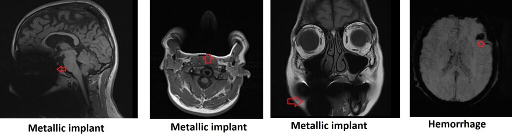
Magnetic susceptibility artifact in MRI refers to distortions or signal voids that can occur due to differences in magnetic susceptibility between different tissues or materials. This artifact is caused by the magnetic field inhomogeneity generated by the magnetic properties of certain materials, such as metallic implants and hemorrhage.
Magnetic susceptibility artifact can cause several imaging artifacts, such as geometric distortion, signal dropout, and blooming artifact. Geometric distortion can cause a misalignment of the image, while signal dropout can cause a loss of signal intensity in the region of interest. Blooming artifact can result in signal spreading from the region of interest into adjacent areas, leading to blurring and loss of detail.
The appearance of magnetic susceptibility artifact depends on the location and type of material causing the artifact. For example, air/tissue interfaces can cause signal voids, while metal implants can cause geometric distortions and signal loss. In some cases, the artifact may appear as a ring or halo around the area of interest.
Here are some strategies to minimize or avoid Magnetic Susceptibility Artifact
Increase bandwidth: Increasing the receiver bandwidth can also help reduce the magnetic susceptibility artifact. A wider bandwidth allows for faster signal acquisition and better compensation for the magnetic field distortions.
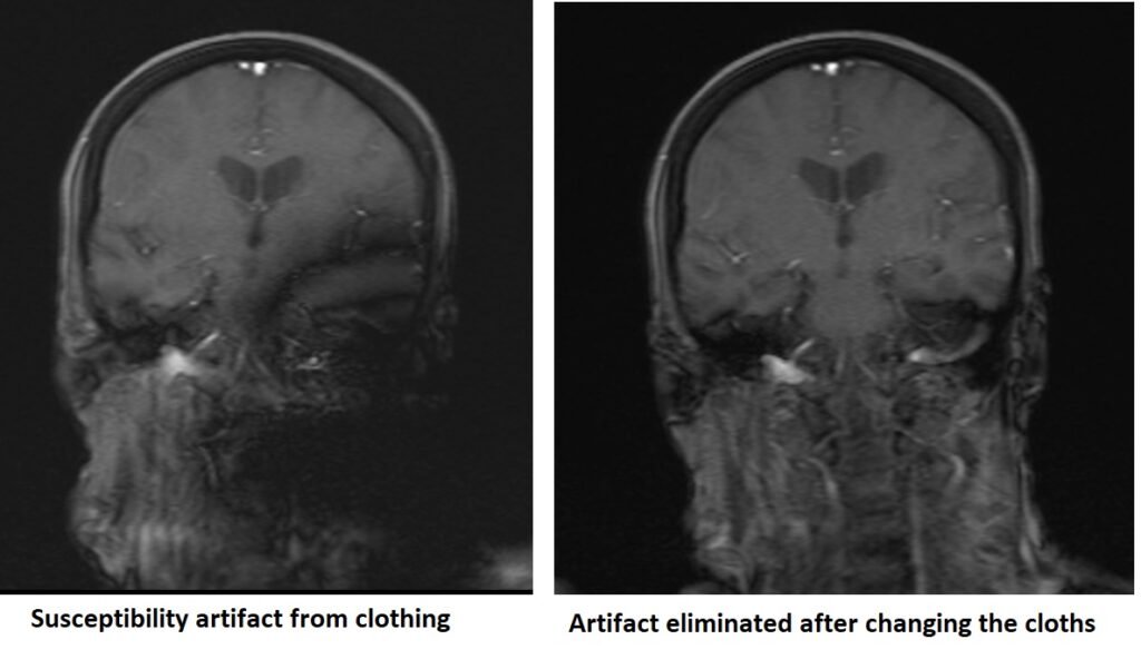
‘Demetallise’ patient thoroughly before the scan: If possible, it is advisable to remove or replace metallic objects or implants before the MRI scan. This helps to eliminate or reduce the source of the magnetic field distortions caused by these materials.
Alter phase and frequency encoding directions: By changing the phase and frequency encoding directions in the MRI acquisition, it is possible to “throw” the artifact away from the region of interest. This can help minimize the impact of the artifact on the critical areas of the image.
Use spin echo sequences rather than gradient echo (GRE): Spin echo sequences are less susceptible to magnetic field variations compared to GRE sequences. By using spin echo sequences, the refocusing of the magnetization can help minimize the effects of magnetic field distortions.
Decrease voxel size/slice thickness: By reducing the voxel size or slice thickness, the magnetic field differences within the imaging volume are minimized, resulting in fewer susceptibility artifacts.
Use dedicated metal artifact reduction sequences (MARS): Specialized sequences, such as Metal Artifact Reduction Sequences (MARS), are designed specifically to minimize the effects of metal implants on image quality. These sequences utilize specific pulse sequences and reconstruction techniques to mitigate the magnetic susceptibility artifact caused by metallic objects.
Use short echo time (TE): Short TE values can reduce the dephasing effects caused by magnetic field variations, leading to improved image quality with less susceptibility artifacts.
Magnetic susceptibility artifact image gallery


References:
- Haacke EM, Brown RW, Thompson MR, Venkatesan R. Magnetic Resonance Imaging: Physical Principles and Sequence Design. John Wiley & Sons; 1999.
- Lauenstein TC, Sharma P, Hughes T, Heberlein K, Tudorascu DL, Martin DR. Evaluation of optimized inversion‐recovery fat‐suppression techniques for T2‐weighted abdominal MRI. Journal of Magnetic Resonance Imaging. 2008 Mar;27(3):623-8.