MRI Navigators
Navigator Echo
Navigator Echo, also known as Prospective Motion Correction (PMC) or Motion Navigator, is a technique used in magnetic resonance imaging (MRI) to mitigate the effects of motion artifacts and improve image quality. Motion, such as patient movement or physiological motion (like breathing or heartbeat), can lead to blurring and distortion in the MRI images. Navigators help to acquire additional information about the motion and allow the MRI system to adjust the imaging parameters in real-time to compensate for the motion, resulting in clearer and more accurate images.
Functioning of Navigators:
- The initial step involves integrating a navigator pulse sequence into the primary imaging sequence. This specific pulse sequence is tailored to excite and receive signals from the navigator Region of Interest (ROI).
- The navigator ROI is typically positioned over an immobile anatomical region or tissue minimally affected by motion. For instance, during abdominal imaging, the navigator ROI could be placed on the diaphragm or within the liver tissue, depending on the selected navigator type.
- Throughout data acquisition, the MRI system constantly monitors signals from the navigator ROI, contrasting them with a reference signal or baseline. Any deviations from the baseline signal indicate patient motion.
- Upon detecting motion, the MRI system promptly adapts the imaging sequence in real-time, which may involve modifying phase encoding steps, adjusting readout gradients, or repeating specific sequence components.
Physics of Navigators:
A navigation sequence is comprised of an intermittent, two-dimensional pulse that stimulates a cluster of spins arranged cylindrically. This is succeeded by a readout gradient aligned with the cylinder’s long axis, capturing a one-dimensional profile of the targeted region. To mitigate signal saturation and abrupt signal intensity shifts in the lungs and liver along the cylinder axis, a modest flip angle of 10 degrees is employed. This intensity change is exploited to determine the diaphragm’s position. The navigator pulse persists for around 20 milliseconds and is executed at intervals of 200 milliseconds. Following the processing of preliminary pre-scan data, a scanning acceptance window is determined, marking the initiation of the actual scan acquisition. In each slice acquisition, the navigation framework detects the diaphragm’s position, allowing imaging exclusively when the diaphragm aligns within the predetermined acceptance window.
The latest generation of scanners offers two distinct types of navigator options:

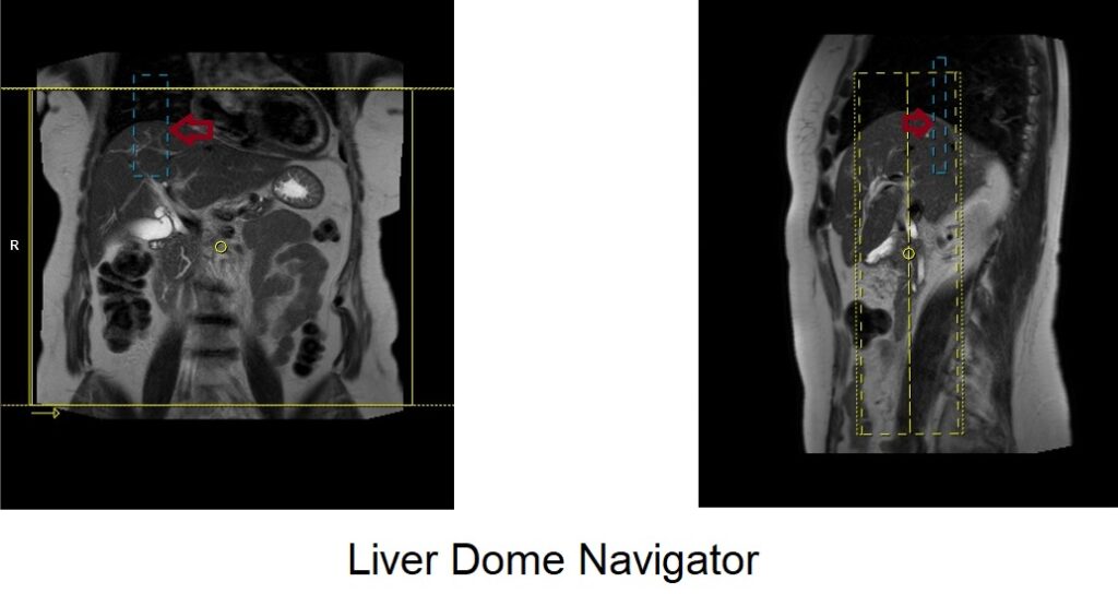
Liver Dome Navigator:
The Liver Dome Navigator necessitates placing a Region of Interest (ROI) over the liver dome (spanning both the liver and chest). The MRI system continuously monitors the ROI’s position as the patient breathes. If the ROI ventures beyond a predefined range, the system adjusts triggering and imaging sequence timing to counteract motion effects. This enhances the acquisition of clear images of the liver and adjacent structures. To ensure precision in Liver Dome Navigator scans, accurate placement of the navigator box is vital. Position it at the midpoint of the right diaphragm dome, with half of the box encompassing the right liver lobe (segment 8) and the remainder over the lungs. Planning should be executed using a non-breath-hold localizer, considering the diaphragm’s downward liver displacement during inhalation, which could otherwise lead to improper slice planning and navigator box positioning.
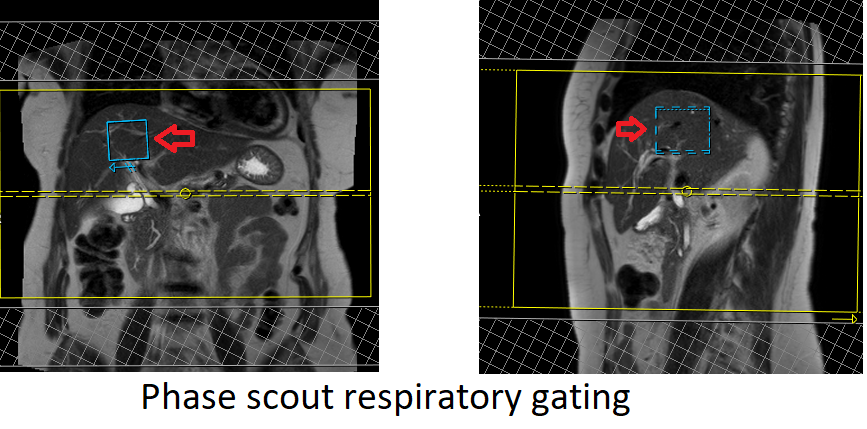
Phase Scout Navigator:
The Phase Scout Navigator is a method to capture a reference image capturing the patient’s respiratory phase. It is primarily used for planning and alignment before actual imaging sequences are executed. This navigator involves acquiring a single low-resolution image (scout image) within a brief timeframe. The acquired image facilitates determining the precise respiratory phase, which subsequently guides the initiation timing of actual imaging sequences. This ensures imaging occurs during a specific respiratory phase, minimizing motion-related artifacts. When employing respiratory gated scans with Phase Scout Navigators, meticulous positioning of the respiratory navigator box within the liver is essential. Ensure that no part of the navigation box extends beyond liver boundaries. Planning should be conducted using a free-breathing localizer to account for the diaphragm’s downward movement during inhalation, preventing inaccuracies in slice planning and navigator box positioning.
References
- Hargreaves BA, Vasanawala SS, Nayak KS, et al. Navigator techniques for respiratory motion correction in MRI. Seminars in Ultrasound, CT, and MRI. 2006;27(1):41-50.
- Madore B. MRI motion correction with prospective acquisition and retrospective image-based navigators. Magnetic Resonance in Medicine. 2010;63(4):971-978.
- Reeder SB, Wintersperger BJ, Dietrich O, et al. Practical approaches to the evaluation of signal-to-noise ratio performance with parallel imaging: application with cardiac imaging and a 32-channel cardiac coil. Magnetic Resonance in Medicine. 2005;54(3):748-754.
- Du YP, Parker DL, Davis WL, Cao G. Reduction of respiratory artifacts in MR imaging of the liver at 3.0 T with 2D navigator echoes. Magnetic Resonance Imaging. 2002;20(1):49-56.
- Seiberlich N, Ehses P, Duerk J, Gilkeson R, Griswold M. Improved radial GRAPPA calibration for real-time free-breathing cardiac imaging. Magnetic Resonance in Medicine. 2010;63(1):19-27.