MRI Velocity Encoding (VENC)
Velocity Encoding (VENC) in Magnetic Resonance Imaging (MRI) is a parameter used to quantitatively assess the velocity of moving fluids within the body, such as blood and cerebrospinal fluid (CSF). VENC defines the maximum velocity that can be accurately measured by the MRI scanner without the occurrence of aliasing artifacts. It is set based on the anticipated fastest flow in the region of interest. In phase-contrast MRI, the VENC value is crucial as it ensures that the phase shifts induced by fluid movement are accurately captured, allowing for precise measurement of the speed and direction of blood and CSF flow. This capability is vital for diagnosing and evaluating conditions related to vascular and cerebrospinal dynamics.
Physics behind the Velocity Encoding (VENC)
Velocity Encoding (VENC) in Magnetic Resonance Imaging (MRI) operates on the principle that moving protons in a magnetic field will experience changes in their phase relative to stationary protons. In phase-contrast MRI, specific gradient pulses are applied to encode the velocity of these moving protons into the phase of the MRI signal. The VENC value is set to calibrate these gradients based on the expected maximum speed of the fluid motion in the imaging field, such as blood or cerebrospinal fluid (CSF). If the fluid moves faster than the set VENC, it causes the phase shift to wrap around, leading to aliasing artifacts in the image. By carefully selecting a VENC close to the highest velocity expected, the MRI system can accurately measure fluid velocities by comparing the phase differences between images obtained with and without these velocity-encoding gradients. This method enables the detailed analysis of fluid dynamics within the body. The unit of measurement for VENC is centimeters per second (cm/s).
Aortic flow is measured with a VENC of 50 cm/s

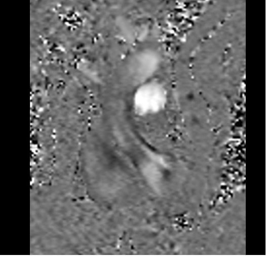
Aortic flow is measured with a VENC of 150 cm/s

Step-by-Step Guide for Radiographers on Calculating and Applying VENC in MRI Flow Sequences
Setting Up the Phase-Contrast Sequence
Phase-contrast MRI sequences utilize gradient pulses to create phase shifts in moving protons, which are directly proportional to their velocity along the direction of the gradient. This technique differentiates between stationary and moving spins by encoding movement into the phase of the MRI signal. The difference in phase between stationary and moving spins can then be used to calculate velocity.
- Initial Setup: The radiographer sets up a basic MRI sequence with specific parameters that are suitable for capturing the flow of interest. This includes selecting the appropriate plane (either through-plane or in-plane) that will intersect the flow.
- VENC Preliminary Estimate: An initial estimate of the VENC is made based on expected physiological velocities in the vessel or area of interest. For example, higher VENC values are used for arteries with high flow rates (like the aorta), and lower values are used for veins or areas with slower flow
Performing a VENC Scout Scan
- Scout Scans: Before the final flow measurement, a series of scout scans (also known as VENC test scans or range-finding scans) are performed. These scans cover a range of VENC values, starting from a low estimate to higher velocities.
- Purpose of Scout Scans: These scans help in identifying the VENC value at which there is no aliasing (where the velocity measurements wrap around and appear falsely low) and yet the sensitivity of the scan is maximized for the velocities encountered.
Analyzing Scout Scan Results
- Reviewing Images: The radiographer and possibly a radiologist review the images from the scout scans to determine the optimal VENC value. This value is the highest velocity that can be measured without aliasing and provides the best contrast for the flow dynamics being studied.
- Selection of Optimal VENC: The optimal VENC is selected based on a balance between avoiding aliasing and maximizing image quality. If aliasing is observed in the preliminary scans, the VENC is adjusted upward; if the images appear too noisy or the low velocities are not well visualized, it may be adjusted downward.
Setting the Final Phase-Contrast Sequence
- Final VENC Setting: Once the optimal VENC value is determined, it is set as the parameter for the final phase-contrast flow sequence.
- Execution of Final Scan: The final detailed phase-contrast sequence is then run using this optimized VENC setting. This sequence will provide quantitative data on the flow velocities, which can be used for clinical assessment and diagnosis.
Quantitative Analysis
- Post-Processing: After the scan, the images are post-processed using software that calculates flow volumes, velocities, and patterns. These quantitative measures are essential for diagnosing conditions like stenosis, valve malfunctions, or shunts.
VENC Scout Conducted for Aortic Through-Plane Flow Analysis

VENC values used for different arteries and CSF
CSF in the Brain: VENC Value: 5-10 cm/s
CSF in the Spinal Cord: VENC Value: 5-10 cm/s
Carotid Artery: VENC Value: Typically around 50-100 cm/s, but this may need to be adjusted based on specific patient conditions.
Aorta: VENC Value: Generally higher, around 130-200 cm/s to account for the high flow velocity through the major artery.
Pulmonary Artery: VENC Value: 60-130 cm/s, tailored to reflect the dynamic nature and the variable flow rates depending on physiological or pathological states.
Pulmonary Vein: VENC Value: 45-70 cm/s, considering the slightly lower flow rates compared to the arterial side of the pulmonary circulation.
Femoral Artery: VENC Value: Similar to the carotid artery, around 50-100 cm/s, reflecting its status as a major conduit of arterial blood to the lower extremities.
Jugular Vein: VENC Value: 30-40 cm/s, appropriate for the venous return flow which is slower than arterial flow.
Superior Vena Cava (SVC) and Inferior Vena Cava (IVC): VENC Value: 30-50 cm/s, suitable for capturing the venous return to the heart without aliasing.
Aortic flow is measured with a VENC of 160 cm/s

Pulmonary flow is measured with a VENC of 110 cm/s
Carotid Artery flow is measured with a VENC of 70 cm/s

CSF flow is measured with a VENC of 6 cm/s

References
- Sarrami-Foroushani, A., Nasr Esfahany, M., Nasiraei Moghaddam, A., Saligheh Rad, H., Firouznia, K., Shakiba, M., Ghanaati, H., Wilkinson, I. D., & Frangi, A. F. (2015). Velocity Measurement in Carotid Artery: Quantitative Comparison of Time-Resolved 3D Phase-Contrast MRI and Image-based Computational Fluid Dynamics. Iranian Journal of Radiology, 12(4), e18286. https://doi.org/10.5812/iranjradiol.18286
- Korbecki, A., Zimny, A., Podgórski, P., Sąsiadek, M., & Bladowska, J. (2019). Imaging of cerebrospinal fluid flow: Fundamentals, techniques, and clinical applications of phase-contrast magnetic resonance imaging. Polish Journal of Radiology, 84, e240–e250. https://doi.org/10.5114/pjr.2019.86881
- Battal, B., Kocaoglu, M., Bulakbasi, N., Husmen, G., Sanal, H. T., & Tayfun, C. (2011). Cerebrospinal fluid flow imaging by using phase-contrast MR technique. British Journal of Radiology, 84(1004), 758-765. https://doi.org/10.1259/bjr/66206791
- Nayak, K. S., Nielsen, J.-F., Bernstein, M. A., Markl, M., Gatehouse, P. D., Botnar, R. M., Saloner, D., Lorenz, C., Wen, H., Hu, B. S., Epstein, F. H., Oshinski, J. N., & Raman, S. V. (2015). Cardiovascular magnetic resonance phase contrast imaging. Journal of Cardiovascular Magnetic Resonance, 17(1), 71. https://doi.org/10.1186/s12968-015-0172-7
- Reiter, U., Reiter, G., & Fuchsjäger, M. (2016). MR phase-contrast imaging in pulmonary hypertension. British Journal of Radiology, 89(1063), 20150995. https://doi.org/10.1259/bjr.20150995