MRI Chemical Shift Artifacts
Chemical shift artifact occurs due to the displacement of the signal, typically along the borders between fat and fluid. Hydrogen atoms in fat and water have different precessional frequencies, resulting in a shift in their recorded locations. At 1.5T, the frequency shift between fat and water is approximately 220 Hz. This discrepancy during frequency encoding leads to mis-mapping of the relative positions of fat and water on the image.
The effect on the image is seen as a dark band on one side and a bright band on the other side of the fat-water or fat-soft tissue interface in spin echo and gradient echo sequences. This artifact is particularly noticeable where fat surrounds organs with high water content, such as the kidney, optic nerve, and liver. It can also occur at the borders between substances with different chemical shifts, such as breast tissue and a silicone implant.

Here are some strategies to minimize or avoid Chemical shift artifact :
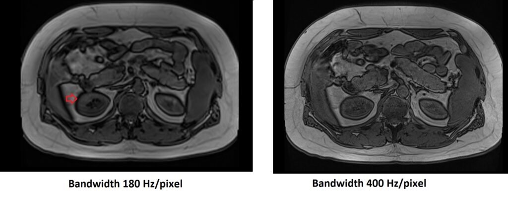
Increase receiver bandwidth: By increasing the receiver bandwidth, a wider range of frequencies can be accepted from each pixel, reducing the shift and minimizing the artifact. However, this approach may result in a reduction in signal-to-noise ratio (SNR).
Use STIR (short tau inversion recovery) or fat saturation sequence: Applying techniques like STIR or fat saturation can effectively remove the fat signal, thereby mitigating the artifact.

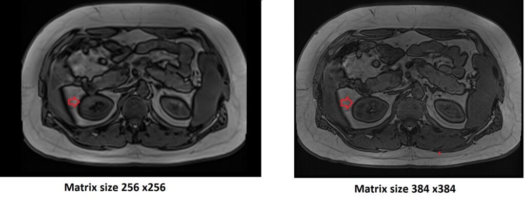
Increase matrix size: Increasing the matrix size can reduce the impact of chemical shift artifact. However, this may also lead to a reduction in SNR.
Swap phase and frequency encoding: Changing the direction of chemical shift by swapping the phase and frequency encoding can help minimize the artifact.
For echo planar imaging (EPI), chemical shift artifact is more prominent in the phase encoding (PE) direction rather than the frequency encoding (FE) direction. In EPI, multiple phase encode lines are collected in a single TR, resulting in a narrower bandwidth per pixel in the PE direction. This can exacerbate the appearance of the chemical shift artifact.
References:
- Reeder SB, Wen Z, Yu H, et al. Multicoil Dixon chemical species separation with an iterative least-squares estimation method. Magn Reson Med. 2004;51(1):35-45.
- Lauenstein TC, Sharma P, Hughes T, et al. Evaluation of optimized inversion-recovery fat-suppression techniques for T2-weighted abdominal MR imaging. J Magn Reson Imaging. 2008;27(6):1448-1454.