3T MRA Brain Anatomy High-resolution 1 mm Slices (Subscription-Based Premium Access)
Explore our advanced MRA brain anatomy library, featuring high-resolution labelled magnetic resonance angiography images of the intracranial arterial system. This specialist module is designed to help users understand the cerebral arteries, circle of Willis, major intracranial branches, vascular territories, and arterial relationships in a clinically realistic imaging format.
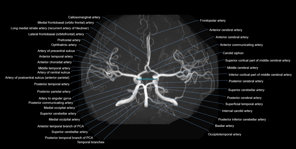
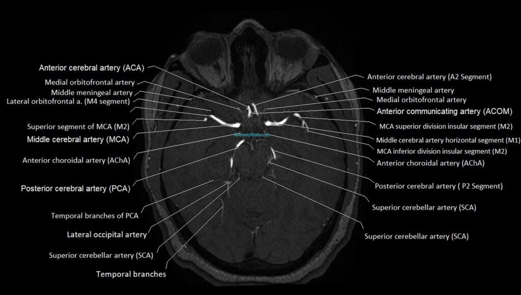
The MRA brain section includes the internal carotid arteries, anterior cerebral arteries, middle cerebral arteries, posterior cerebral arteries, anterior communicating artery, posterior communicating arteries, basilar artery, vertebral arteries, superior cerebellar arteries, anterior inferior cerebellar arteries, posterior inferior cerebellar arteries, ophthalmic arteries, lenticulostriate arteries, and major cortical branches.
The vascular anatomy is manually labelled with careful attention to branch origin, course, and segmental anatomy. This allows users to learn important structures such as ACA A1–A3 segments, MCA M1–M4 segments, PCA P1–P3 segments, carotid siphon, basilar tip, vertebrobasilar junction, and circle of Willis variants.
The MRA images are built into an interactive scroller and viewer, allowing users to review the arterial anatomy in a way that reflects clinical vascular imaging practice. The interface is designed for desktop and mobile use, with full-screen viewing for detailed study.
Voice and text search allow users to quickly find specific arteries and access detailed descriptions of their vascular territory, imaging appearance, and anatomical relationships. This supports learning for stroke imaging, aneurysm localisation, vascular malformations, surgical planning, and interventional radiology.
The zoom function allows high-detail review of small arterial branches and vascular junctions without compromising image quality.
The MRA brain anatomy module is supported by dynamic quizzes that generate new questions in every session, helping users build lasting knowledge of cerebral vascular anatomy and intracranial arterial relationships.
MRA Brain Axial 1mm Labelled Image(sample)
MRA Brain MIP Labelled Image(sample)