DESS /FADE MRI
DESS (Double Echo Steady State) is a specialized MRI sequence primarily employed in musculoskeletal imaging, providing high-resolution images of cartilage. It proves particularly valuable in assessing articular cartilage and detecting cartilage lesions.
The three-dimensional double-echo steady-state (3D-DESS) sequence utilizes a 3D gradient echo technique, allowing for the simultaneous acquisition of two distinct steady-state free precession (SSFP) echoes with entirely different contrasts: the FISP sequence and the PSIF sequence. These echoes are subsequently combined using a sum of squares calculation. While the PSIF component imparts high T2 contrast, the FISP portion generates morphological images predominantly influenced by the T1/T2 ratio. This amalgamated data results in a T2*-weighted image, significantly enhancing sensitivity and specificity in the imaging of cartilage and synovial fluid
MRI Image Appearance of DESS MRI
The simplest method to recognize DESS images is by looking for fluid-filled regions within the body, like cerebrospinal fluid in brain ventricles and the spinal canal, synovial fluid in joints, or any other abnormal fluid accumulations. Typically, fluids exhibit a bright appearance in DESS images, while muscles and fat generally appear as gray. In instances of fat-saturated DESS, both muscles and fat may appear dark.
DESS MRI Applications
Orthopedic Imaging: DESS is widely used in the evaluation of joints, especially the knee, ankle, and wrist. It’s particularly beneficial for visualizing cartilage, cartilage lesions, and other related pathologies.
Cartilage Mapping: Due to its high-resolution capabilities, DESS can be used for cartilage mapping, which is a technique to quantitatively evaluate cartilage thickness and volume.
Tissues and their DESS appearance
Bone marrow – dark
Muscles – bright gray
Fat – gray
Moving blood– bright
Spinal cord – gray
Cartilage – bright
Fluids – bright
Bone – dark
Air – dark
Tissues and their fat saturated DESS appearance
Bone marrow – dark
Muscles – dark gray
Fat – dark gray (darker than muscle)
Moving blood– bright
Spinal cord – gray
Cartilage – bright
Fluids – bright
Bone – dark
Air – dark
Use
- Very useful for joints imaging (fat saturated DESS in knee, hips, ankle, shoulder, wrist and elbow)
- Very useful for cartilage mapping
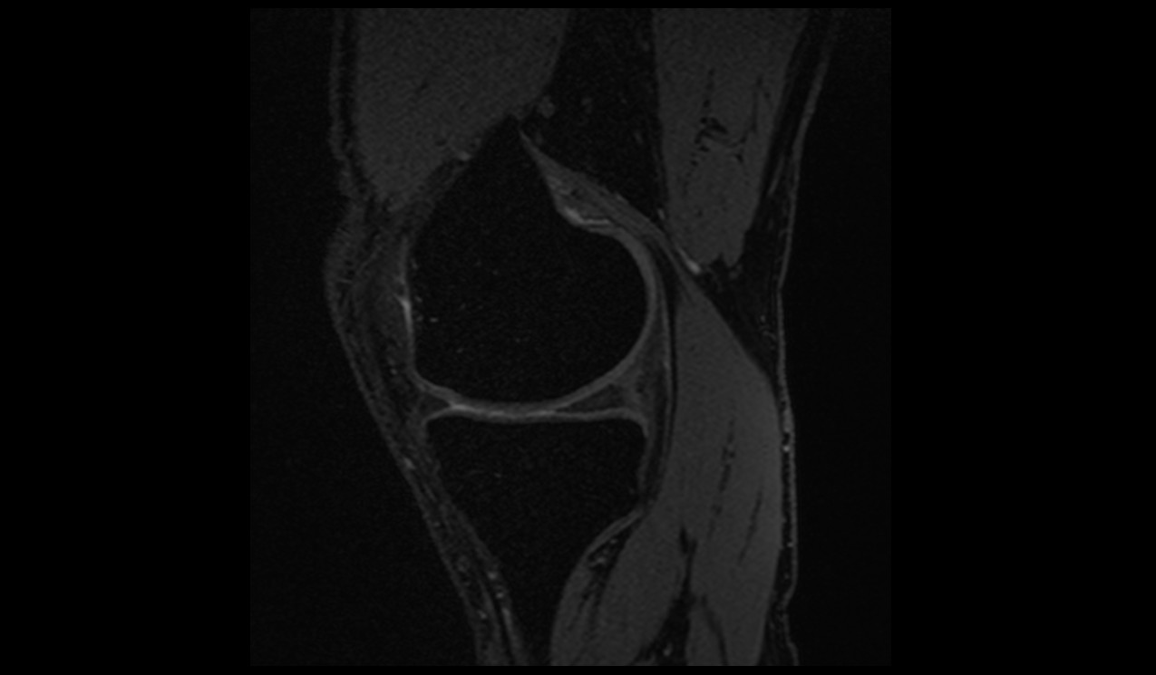
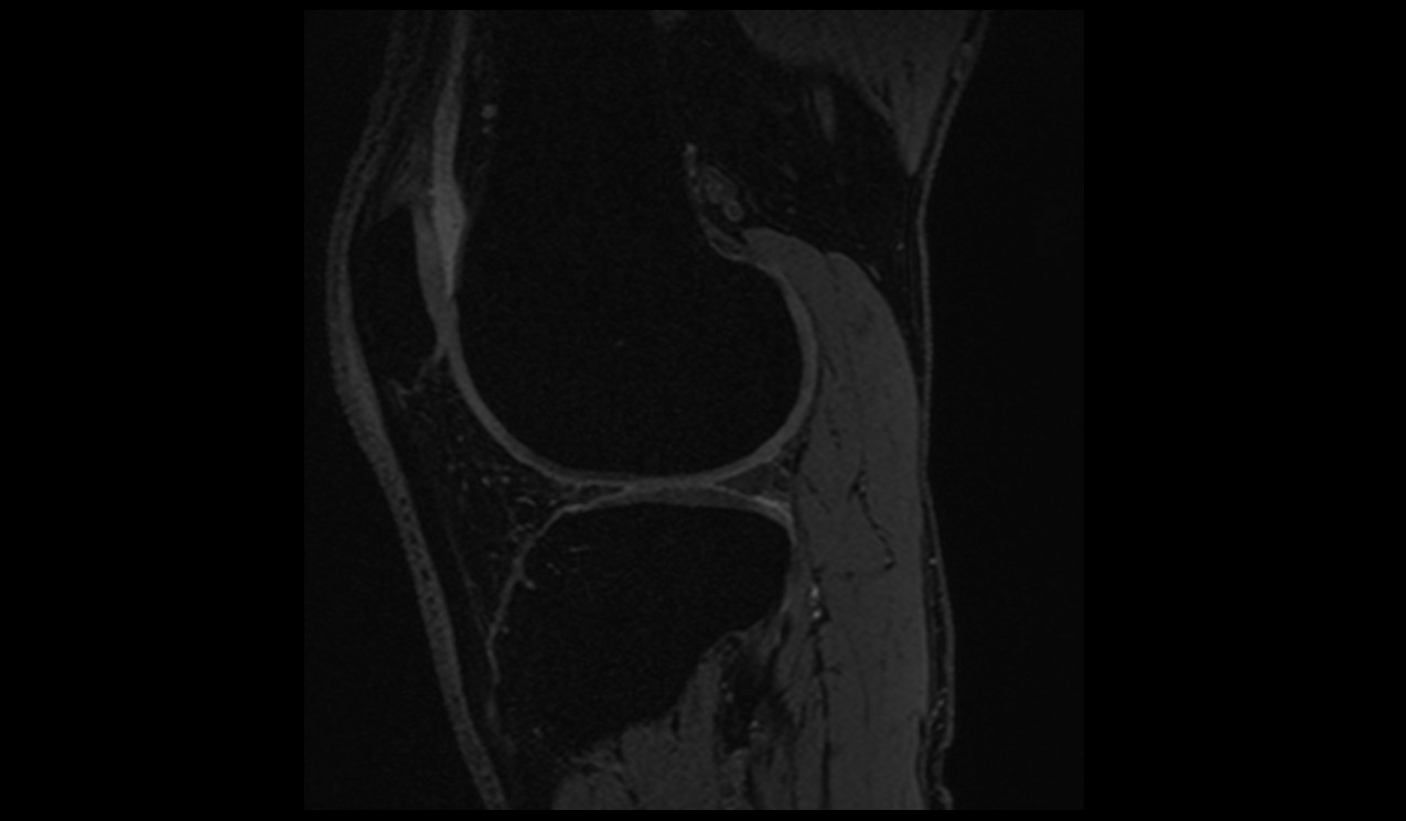
DESS sagittal image of Knee
DESS coronal image of Shoulder
