Pectoralis Muscle MRI
Indications for pectoralis muscle mri
- Pectoralis major ruptures
- Pectoralis major injuries
- Pectoralis muscle tumor
- Pectoralis muscle tumor
- chest wall tumor
- Ligament tear
Contraindications
- Any electrically, magnetically or mechanically activated implant (e.g. cardiac pacemaker, insulin pump biostimulator, neurostimulator, cochlear implant, and hearing aids)
- Intracranial aneurysm clips (unless made of titanium)
- Pregnancy (risk vs benefit ratio to be assessed)
- Ferromagnetic surgical clips or staples
- Metallic foreign body in the eye
- Metal shrapnel or bullet
Patient preparation for pectoralis muscle mri
- A satisfactory written consent form must be taken from the patient before entering the scanner room
- Ask the patient to remove all metal objects including keys, coins, wallet, cards with magnetic strips, jewellery, hearing aid and hairpins
- If possible provide a chaperone for claustrophobic patients (e.g. relative or staff )
- Offer earplugs or headphones, possibly with music for extra comfort
- Explain the procedure to the patient
- Instruct the patient to keep still
- Note the weight of the patient
Positioning for MRI pectoralis muscle mri
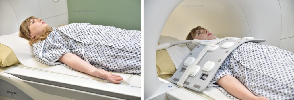
- Position patient in the supine position with head pointing towards the magnet (head first supine).
- Position the patient off-center over the spine coil as demonstrated, and then place the large flexible coil over the chest. Adjust the flexible coil so that it is positioned more towards the affected side.
- Securely tighten the body coil using straps to prevent respiratory artefacts
- The patient may be given a pillow and cushions under their legs for extra comfort
- Centre the laser beam localiser over the sternoclavicular joint
- Register the patient on the scanner as 'head first supine'
Recommended Pectoralis Muscle MRI Protocols and Planning
Localiser 1
A three-plane localizer must be taken in the beginning to localize and plan the sequences. Localizers are usually less than 25 seconds. T2-weighted low-resolution scans are used for this purpose.

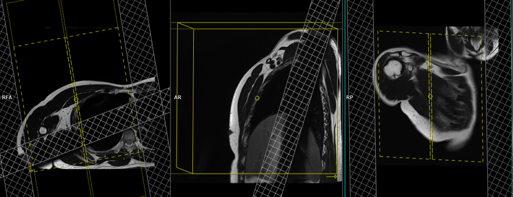
Localiser 2
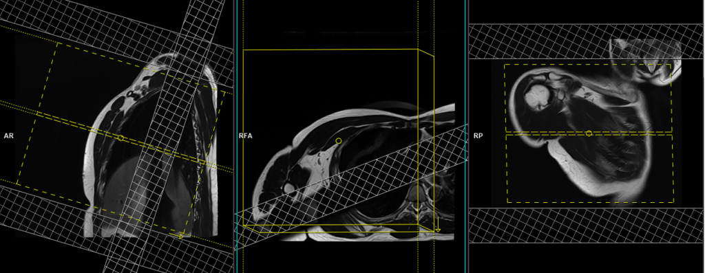
Plan the coronal and sagittal localizers based on the axial plane. Angle the planning block parallel to the pectoralis muscle for the coronal localizer, and angle it perpendicular to the pectoralis muscle for the sagittal localizer. For an additional axial localizer, position the block perpendicular to the chest on the sagittal plane.

T2 HASTE\T2 TSE breath hold axial 4mm SFOV
Plan the axial slices in the coronal plane and position the block horizontally across the pectoralis muscle. Check the positioning block in the other two planes. An appropriate angle must be used in the sagittal plane, perpendicular to the chest wall. Ensure that the slices are sufficient to cover the entire pectoralis muscle, from the top of the acromioclavicular joint to 2 inches below the xiphoid sternum. Adding saturation bands over the chest will help reduce ghosting artifacts caused by breathing. The phase direction can be anteroposterior or right to left, with sufficient oversampling to avoid wrap-around and ghosting artifacts originating from the chest
Parameters T2 tse BH
TR 4000-5000 | TE 90 | FLIP 150 | NXA 1 | SLICE 4 MM | MATRIX 224×224 | FOV 250 | PHASE A>P | OVERSAMPLE 50% | BH YES |
T2 HASTE\T2 TSE fat saturated breath hold axial 4mm SFOV
Plan the axial slices in the coronal plane and position the block horizontally across the pectoralis muscle. Check the positioning block in the other two planes. An appropriate angle must be used in the sagittal plane, perpendicular to the chest wall. Ensure that the slices are sufficient to cover the entire pectoralis muscle, from the top of the acromioclavicular joint to 2 inches below the xiphoid sternum. Adding saturation bands over the chest will help reduce ghosting artifacts caused by breathing. The phase direction can be anteroposterior or right to left, with sufficient oversampling to avoid wrap-around and ghosting artifacts originating from the chest
Parameters T2 tse fat saturated BH
TR 6000-8000 | TE 90 | fatsat ON | NXA 1 | SLICE 4 MM | MATRIX 208×208 | FOV 250 | PHASE A>P | OVERSAMPLE 30% | IPAT ON |
T1 VIBE DIXON(In-phase and water sat) breath hold axial 4mm SFOV
Plan the axial slices on the coronal plane and angle the positioning block horizontally across the scapular blade. Check the positioning block in the other two planes. An appropriate angle must be used in the sagittal plane, perpendicular to the scapular blade. Slices must be sufficient to cover the whole scapula from the top of the acromioclavicular joint to 1 cm below the inferior angle of the scapula. Adding saturation bands over the chest will help reduce ghosting artifacts from breathing. The phase direction must be anteroposterior with sufficient oversampling to avoid wrap-around and ghosting artifacts from the chest.
Parameters
TR 7-8 | TE 2.39 4.77 | FLIP 10 | NXA 1 | SLICE 4 MM | MATRIX 256×224 | FOV 250 | PHASE R>L | OVERSAMPLE 50% | BH YES |
T2 HASTE\T2 TSE fat saturated breath hold coronal 4mm SFOV
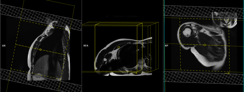
Plan the coronal slices based on the axial plane and align the planning block parallel to the pectoralis muscle. Verify the planning block in the other two planes. In the sagittal plane, employ an appropriate angle parallel to the chest wall. Ensure that the slices adequately cover the entire pectoralis muscle, from the anterior skin margin to the posterior mid-humeral head. To minimize ghosting artifacts caused by breathing, consider incorporating an oblique saturation band over the chest. For the phase direction, utilize right to left with sufficient oversampling to prevent wrap-around and ghosting artifacts originating from the chest.
Parameters
TR 6000-8000 | TE 90 | fatsat ON | NXA 1 | SLICE 4 MM | MATRIX 208×208 | FOV 250 | PHASE R>L | OVERSAMPLE 70% | IPAT ON |
T1 VIBE DIXON(In-phase and water sat) breath hold coronal 4mm SFOV
Plan the coronal slices based on the axial plane and align the planning block parallel to the pectoralis muscle. Verify the planning block in the other two planes. In the sagittal plane, employ an appropriate angle parallel to the chest wall. Ensure that the slices adequately cover the entire pectoralis muscle, from the anterior skin margin to the posterior mid-humeral head. To minimize ghosting artifacts caused by breathing, consider incorporating an oblique saturation band over the chest. For the phase direction, utilize right to left with sufficient oversampling to prevent wrap-around and ghosting artifacts originating from the chest.

Parameters
TR 7-8 | TE 2.39 4.77 | FLIP 10 | NXA 1 | SLICE 4 MM | MATRIX 256×224 | FOV 250 | PHASE R>L | OVERSAMPLE 70% | BH YES |
T2 HASTE\T2 TSE breath hold sagittal 4mm SFOV
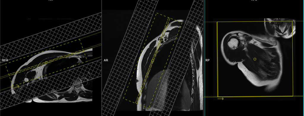
Plan the sagittal slices on the axial plane and position the block perpendicular to the pectoralis muscle. Verify the positioning block in the other two planes. Provide an appropriate angle in the coronal plane, running vertically across the pectoralis muscle. Ensure that the slices cover the pectoralis muscle, extending from the humeral head to the mid-sternum. To minimize ghosting artifacts caused by breathing, incorporate an oblique saturation band over the chest. Choose the phase direction as either anteroposterior or head to feet, with ample oversampling to prevent wrap-around and ghosting artifacts.
Parameters
TR 4000-5000 | TE 90 | FLIP 150 | NXA 1 | SLICE 4 MM | MATRIX 224×224 | FOV 250 | PHASE A>P | OVERSAMPLE 50% | BH YES |
Optional Scans
DWI epi 3 scan trace axial 3mm free breathing
Plan the axial epi DWI slices in the coronal plane and position the block horizontally across the pectoralis muscle. Check the positioning block in the other two planes. An appropriate angle must be used in the sagittal plane, perpendicular to the chest wall. Ensure that the slices are sufficient to cover the entire pectoralis muscle, from the top of the acromioclavicular joint to 2 inches below the xiphoid sternum. Adding saturation bands over the chest will help reduce ghosting artifacts caused by breathing. The phase direction can be anteroposterior or right to left, with sufficient oversampling to avoid wrap-around and ghosting artifacts originating from the chest
Parameters
TR 6000-7000 | TE 90 | IPAT ON | NEX 3 5 8 | SLICE 3 MM | MATRIX 192X192 | FOV 200-250 | PHASE R>L | GAP 10% | B VALUE 0 |