FLAIR vs STIR MRI
T2 FLAIR MRI
FLAIR MRI, which stands for Fluid-Attenuated Inversion Recovery Magnetic Resonance Imaging, is a special type of MRI sequence used predominantly in neurological imaging to enhance the visibility of lesions and abnormalities in the brain and spinal cord, particularly those associated with demyelinating diseases like Multiple Sclerosis. It suppresses the signal from cerebrospinal fluid (CSF), making abnormalities in the adjacent white matter more conspicuous.
The key feature of FLAIR MRI is its inversion recovery pulse sequence, which involves inverting the magnetization of the water molecules in the body before acquiring the images. This inversion helps nullify the signal from CSF and other fluids, making them appear dark on the resulting images.
Typical inversion times (TI) used in FLAIR MRI sequences may vary slightly depending on the scanner manufacturer and protocols, but common TI values include around 2,000 to 2,500 milliseconds.
What are we looking for in FLAIR images?
In FLAIR images, radiologists primarily search for abnormalities or lesions within the brain tissue. Because it is sensitive to changes in water content, FLAIR is effective for detecting:
- Multiple Sclerosis (MS) lesions: Appearing as hyperintense (bright) regions in the white matter.
- Infarcts or stroke lesions: It helps visualize areas of the brain that have suffered from a lack of blood supply.
- Brain tumors : Brain tumors such as gliomas can be seen more distinctly.
- Infections and inflammation: FLAIR can help detect abnormalities related to infections or inflammatory processes in the brain.
- White matter diseases: FLAIR is effective in detecting various white matter diseases and abnormalities related to demyelination.
Explore further about FLAIR MRI in our dedicated FLAIR MRI section.
STIR MRI
STIR MRI, standing for Short TI Inversion Recovery Magnetic Resonance Imaging, is a unique type of MRI sequence primarily used in musculoskeletal and body imaging to amplify the visibility of lesions and abnormalities within the body structures, especially those obscured by fat. It is renowned for suppressing the signal from fat, enabling abnormalities adjacent to or within fatty tissues to be more conspicuous.
The principal feature of STIR MRI is its short inversion recovery pulse sequence, where the magnetization of the hydrogen atoms primarily associated with fat is inverted before acquiring the images. This inversion helps to nullify the signal from fat, making fatty tissues appear dark on the resulting images.
STIR MRI, much like FLAIR MRI, utilizes specific inversion times (TI) to achieve its contrast objectives. The choice of TI values may vary depending on the scanner manufacturer and clinical protocols, but typically falls within the range of 130 to 180 milliseconds.
What are we looking for in STIR images?
In STIR images, radiologists primarily search for pathologies and abnormalities that can be masked by fat. Due to its sensitivity to water content and its capability to suppress fat signal, STIR is effective for detecting:
- Musculoskeletal Injuries: Injuries to tendons, ligaments, and muscles can be more discernible on STIR images.
- Bone Marrow Pathologies: Lesions or abnormalities in the bone marrow are more conspicuous as the fat signal is suppressed.
- Edema and Inflammation: Both appear hyperintense (bright) on STIR images, making them useful for detecting inflammatory conditions and post-traumatic changes.
- Spinal Pathologies: Lesions or abnormalities in or near the spine, which might be obscured by fatty tissues, can be more evident.
- Tumors and Masses: While many tumors have a fatty component, the non-fatty parts of the tumor or associated edema can be seen more clearly on STIR.
FLAIR and STIR Image Appearance of Various Structures in the Brain
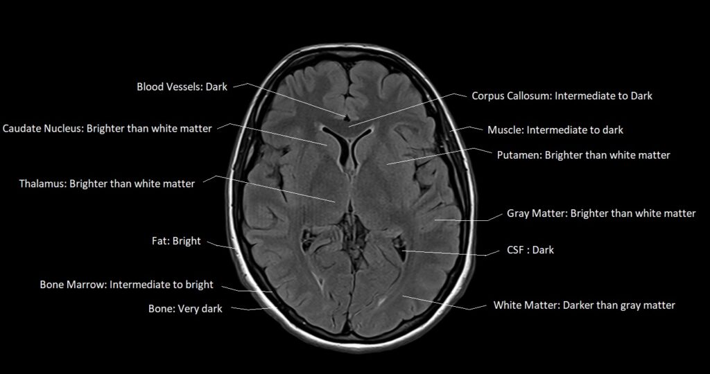
FLAIR appearance of the brain
- Fat: Bright
- Muscle: Intermediate to dark
- CSF : Dark
- White Matter: Darker than gray matter
- Gray Matter: Brighter than white matter
- Bone: Very Dark (low signal)
- Bone Marrow: Variable but often intermediate to bright
- Blood Vessels: Mostly Dark, Depending on flow characteristics, can be bright or dark
- Pituitary Gland: Intermediate
- Choroid Plexus: Intermediate to bright
- Cerebellum (consists of both white and gray matter): Gray matter brighter than white matter
- Brain Stem: Intermediate
- Sinuses: Dark (filled with air)
- Thalamus: Brighter than white matter
- Putamen: Brighter than white matter
- Pineal Gland: Intermediate
- Hippocampus: Brighter than white matter
- Corpus Callosum: Intermediate to Dark
- Caudate Nucleus: Brighter than white matter
STIR appearance of the brain
- Fat: Dark (Due to fat suppression)
- Muscle: Intermediate
- CSF : Bright
- White Matter: Intermediate to Dark
- Gray Matter: Intermediate to Bright
- Bone: Very Dark
- Bone Marrow: Dark (Due to fat suppression)
- Blood Vessels: Mostly Dark, Depending on flow characteristics, can be bright or dark
- Pituitary Gland: Intermediate.
- Choroid Plexus: Intermediate to bright
- Cerebellum (consists of both white and gray matter): Gray matter brighter than white matter
- Brain Stem: Intermediate to Dark
- Sinuses: Dark (filled with air)
- Thalamus: Intermediate to bright, similar to gray matter
- Putamen: Intermediate to bright, similar to gray matter
- Pineal Gland: Intermediate
- Hippocampus: Intermediate to bright, similar to gray matter
- Corpus Callosum: Intermediate to Dark
- Caudate Nucleus: Intermediate to bright, similar to gray matter
FLAIR MRI Image of the Brain
STIR MRI Image of the Brain

FLAIR and STIR Image Appearance of Various Structures in the Spine
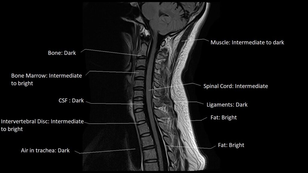
FLAI appearance of the Cervicle spine
- Muscle: Intermediate to dark
- CSF : Dark.
- Bone: Very Dark (low signal).
- Bone Marrow: Intermediate to bright.
- Blood Vessels: Mostly Dark, Depending on flow characteristics, can be bright or dark.
- Spinal Cord: Intermediate signal
- Fat: Bright
- Intervertebral Disc: Nucleus pulposus and annulus bright.
- Ligaments: Intermediate to dark.
- Nerve Roots: Intermediate signal
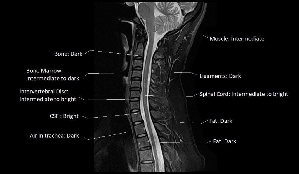
STIR appearance of the Cervicle spine
- Muscle: Intermediate
- CSF : Bright
- Bone: Very Dark
- Bone Marrow: Intermediate to dark
- Blood Vessels: Mostly Dark, Depending on flow characteristics, can be bright or dark
- Spinal Cord: Intermediate to bright
- Fat: Dark
- Intervertebral Disc: Nucleus pulposus brighter, annulus intermediate
- Ligaments: Dark
- Nerve Roots: Intermediate signal
FLAIR MRI Image of the Cervical Spine
STIR MRI Image of the Cervical Spine
References
- Jack, C. R., O’Brien, P. C., Rettman, D. W., et al. (2005). FLAIR MRI in Alzheimer’s disease: An exploration of the in vivo correlates of regional amyloid deposition. Journal of Magnetic Resonance Imaging, 22(6), 849-856.
- Barkhof, F., & Van Walderveen, M. A. (2001). Characterization of tissue damage in multiple sclerosis by nuclear magnetic resonance. Philosophical Transactions of the Royal Society of London. Series B: Biological Sciences, 356(1411), 1653-1663.
- Filippi, M., Rocca, M. A., & Barkhof, F. (2012). Imaging in multiple sclerosis. Neurotherapeutics, 9(1), 43-53.
- Kozak, L. R., White, M. L., O’Malley, J. P., & Haider, M. A. (2012). Magnetic resonance imaging of the musculoskeletal system: a clinical approach. Lippincott Williams & Wilkins.
- Müller M. Short TI inversion recovery (STIR). Magnetic Resonance in Medicine. 1990;14(2):277-295.
- De Coene B., Hajnal J.V., Gatehouse P., Longmore D.B., White S.J., Oatridge A., Pennock J.M., Young I.R., Bydder G.M. (1992). MR of the Brain Using Short Inversion Time Inversion-Recovery Sequences. AJNR. American Journal of Neuroradiology, 13(6), 1555-1564.