MRI Slice Thickness
Slice Thickness
In MRI (Magnetic Resonance Imaging), Slice Thickness refers to the thickness of the individual image slices acquired during the scanning process. It represents the distance between the top and bottom surfaces of the slice or the depth of the section being imaged.
Slice Thickness is typically measured in millimetres and is an essential parameter in determining the level of anatomical detail captured in the image. Thinner slice thickness provides higher spatial resolution and allows for more detailed visualization of structures within the slice. On the other hand, thicker slices can cover a larger volume of tissue but may result in lower spatial resolution and potential loss of fine details.
The selection of an appropriate slice thickness depends on several factors, including the clinical indication, anatomical region of interest, and the trade-offs between resolution and scanning time. For example, in neuroimaging, thinner slices (e.g., 3-4 mm) are often preferred to visualize fine brain structures. In abdominal imaging, thicker slices (e.g., 4-7 mm) may be used to cover larger areas of interest or to balance the need for imaging speed with sufficient anatomical coverage.
PELVIS IMAGE WITH 8MM SLICE THICKNESS

PELVIS IMAGE WITH 3MM SLICE THICKNESS

Slice Thickness and SNR
The SNR (Signal-to-Noise Ratio) in magnetic resonance imaging (MRI) can be influenced by the thickness of the image slices. SNR measures the strength of the signal relative to the level of noise in the image, and a higher SNR generally corresponds to better image quality and improved diagnostic accuracy.
Thinner slices in MRI provide higher spatial resolution and minimize the partial volume effect, which occurs when a single voxel contains multiple tissue types. However, thinner slices tend to have a lower SNR because the signal is distributed over a smaller volume. This can result in a relatively higher level of noise in the image, which may impact the clarity of details.
Thicker slices in MRI can yield a higher SNR since the signal is acquired from a larger volume. This increased volume contributes to a stronger signal, resulting in a higher SNR. However, thicker slices typically have poorer spatial resolution and are more susceptible to the partial volume effect, where details may be blurred or lost due to the inclusion of multiple tissue types within a single voxel.
Slice Thickness 3mm

Slice Thickness 5mm

How to Manipulate Slice Thickness in MRI
Slice thickness and matrix size
Increasing the slice thickness in MRI will result in larger voxel sizes, which are determined by the field of view and the slice thickness. With larger voxels, there is improved SNR since the signal is collected over a larger volume. However, larger voxels also lead to decreased spatial resolution and increased partial volume effect, where different tissues within the voxel mix together.
To achieve an optimal balance between SNR, spatial resolution, and partial volume effect, it is advisable to increase the matrix size, which helps minimize the voxel size. By increasing the matrix size, the spatial resolution can be improved, and the partial volume effect can be reduced.
For example, let’s consider a brain scan with a field of view (FOV) of 200 x 200 mm, a slice thickness of 3 mm, and a matrix size of 288 x 288. If you decide to change the slice thickness to 5 mm, it is recommended to increase the matrix size to 320 x 320 to achieve optimal spatial resolution and SNR.
By adjusting the matrix size along with the change in slice thickness, you can maintain a balance between SNR, spatial resolution, and partial volume effect, ensuring that the acquired images provide the desired image quality and diagnostic information.
Slice thickness 3mm with matrix size 288X288

Slice thickness 5mm with matrix size 320X320

Slice Thickness and NEX (Number of Excitations)
The slice thickness and NEX (number of excitations) in MRI are related, particularly in terms of their impact on the SNR (Signal-to-Noise Ratio) and scan time. By adjusting these parameters, you can achieve an optimum balance between SNR and scan efficiency.
Increasing the slice thickness in MRI tends to improve the SNR because the signal is acquired over a larger volume. This can be advantageous when higher SNR is desired for diagnostic purposes. However, larger slice thickness may result in reduced spatial resolution and increased partial volume effect.
To reduce the SNR to an optimum level while maintaining scan efficiency, you can decrease the NEX (averages). NEX refers to the number of times each slice is acquired and averaged to improve the SNR. By decreasing the NEX, the SNR is reduced, which can be beneficial in situations where excessively high SNR is not necessary.
By reducing the NEX, you can also reduce the scan time since each acquisition takes less time. This can help improve patient comfort and throughput.
For example, let’s consider a brain scan with a field of view (FOV) of 200 x 200 mm, a slice thickness of 3 mm, a matrix size of 288 x 288, and an NEX of 2. If you decide to change the slice thickness to 5 mm, it is recommended to decrease the NEX to 1 to achieve an optimal balance of SNR and scan efficiency.
SLICE THICKNESS 3MM

NEX 2
SLICE THICKNESS 5MM

NEX 1
Slice thickness and FOV (field of view)
The slice thickness and FOV (field of view) in MRI are interconnected and can be adjusted to optimize the SNR (Signal-to-Noise Ratio), spatial resolution, and image quality. By modifying these parameters, you can achieve an optimal balance between them.
Increasing the slice thickness in MRI tends to improve the SNR since the signal is acquired over a larger volume. This can be advantageous when higher SNR is desired for diagnostic purposes. However, larger slice thickness may result in reduced spatial resolution and increased partial volume effect.
To reduce the SNR to an optimum level while improving spatial resolution, you can decrease the FOV. The FOV refers to the physical size of the imaging area covered by the MRI scan. By reducing the FOV, the voxel size is reduced, which can enhance spatial resolution and minimize the partial volume effect.
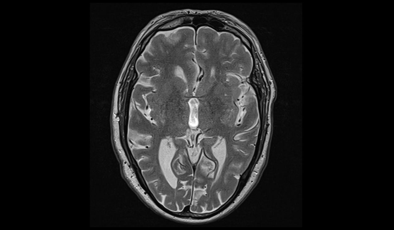
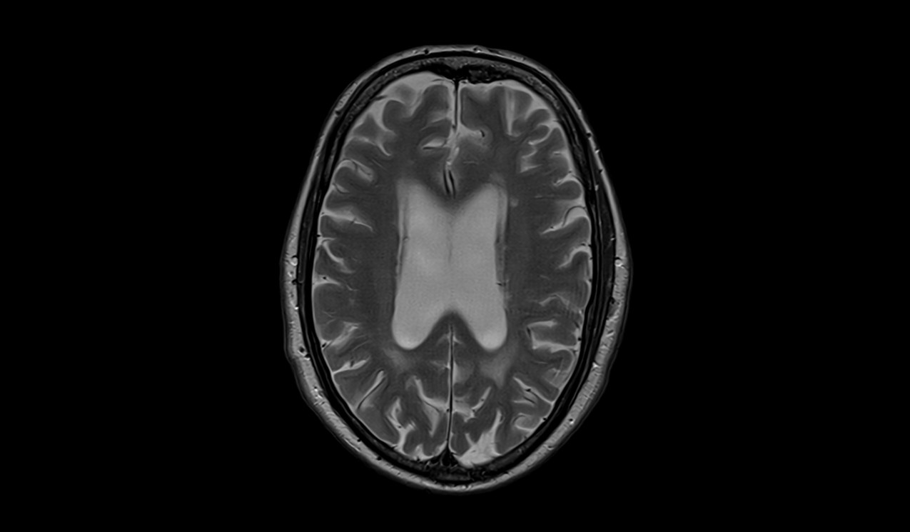
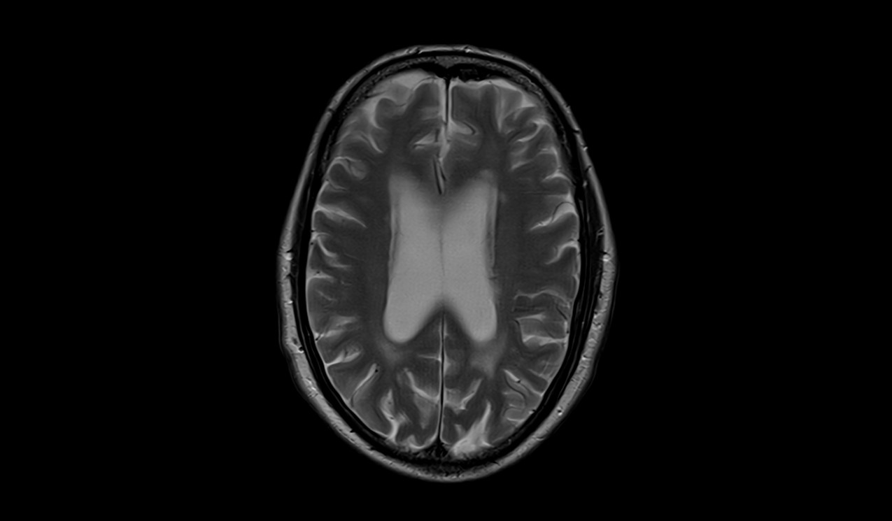
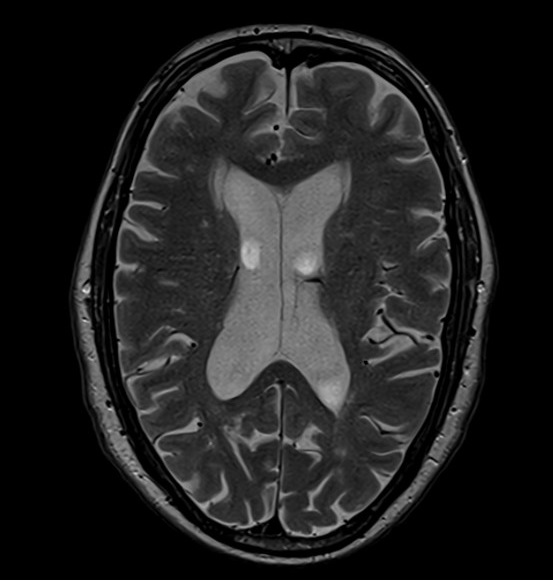
For example, let’s consider a brain scan with an initial FOV of 250 x 250 mm, a slice thickness of 3 mm, and a matrix size of 288 x 288. If you decide to change the slice thickness to 5 mm, it is recommended to decrease the FOV to 200 x 200 mm to achieve an optimal balance of SNR and spatial resolution.
SLICE THICKNESS 3MM

FOV 250MM
SLICE THICKNESS 5MM

FOV 200MM
References
- Vlaardingerbroek, M. T., den Harder, J., van der Jagt, P. K. N., Bovée, W. M. J., Kouwenhoven, M., & Webb, A. G. (2008). On the effect of slice thickness on susceptibility contrast imaging. Magnetic Resonance in Medicine, 60(2), 352–358.
- Reiser, M. F., & Semmler, W. (Eds.). (2007). Magnetic Resonance Tomography. Springer Science & Business Media.
- Haacke, E. M., Brown, R. W., Thompson, M. R., & Venkatesan, R. (2014). Magnetic resonance imaging: Physical principles and sequence design. John Wiley & Sons.
- Pipe, J. G., & Menon, P. (1999). Sampling density compensation in MRI: Rationale and an iterative numerical solution. Magnetic Resonance in Medicine, 41(1), 179-186.
- Walsh, D. O., Gmitro, A. F., & Marcellin, M. W. (2000). Adaptive reconstruction of phased array MR imagery. Magnetic Resonance in Medicine, 43(5), 682-690.
- Lustig, M., Donoho, D., & Pauly, J. M. (2007). Sparse MRI: The application of compressed sensing for rapid MR imaging. Magnetic Resonance in Medicine, 58(6), 1182-1195.
- Truong, T. K., Chen, F., Kim, S. E., & Chang, G. J. (2017). Reducing patient motion in functional MRI using discrete affine transform estimation. Magnetic Resonance in Medicine, 77(5), 1897-1905.
- Nishimura, D. G. (2010). Principles of magnetic resonance imaging: Physics concepts, pulse sequences, & biomedical applications. Stanford University.
