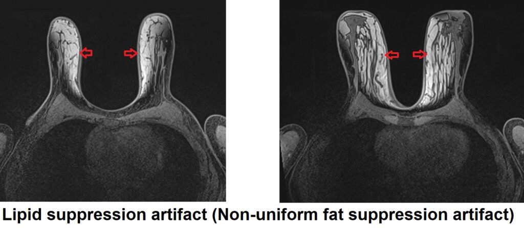
Lipid Suppression Artifact (Fat Suppression Artifact)
Lipid suppression artifact in MRI refers to the incomplete suppression of the signal from fat, resulting in unwanted bright areas in images where fat should be suppressed. This artifact can occur due to several factors, including incorrect pulse sequence parameters, suboptimal shim, and technical issues with the MRI system.
The appearance of lipid suppression artifact in MRI images is usually characterized by bright areas or spots in regions where fat suppression should have occurred. These bright areas can interfere with the diagnosis and interpretation of images, especially in regions where accurate differentiation between fat and other tissues is critical, such as in the musculoskeletal system or breast.
Here are some strategies to minimize or avoid Lipid suppression /Non-uniform fat suppression artifact :
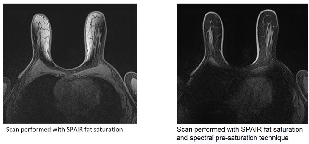
Spectral pre-saturation: Employ spectral pre-saturation techniques to suppress lipid signal. This involves applying selective RF pulses at the resonance frequency of lipid protons before the imaging sequence to reduce their signal. Spectral pre-saturation is effective in reducing lipid artifacts in certain sequences, such as fast spin echo (FSE) or gradient echo (GRE) sequences.
Short tau inversion recovery (STIR): Utilize STIR sequences that null the signal from fat by employing a relatively long inversion time (TI). By nulling the fat signal, STIR sequences can effectively suppress lipid artifacts. However, it’s important to note that STIR sequences also null other signals, so they may not be suitable for all imaging purposes.

Dixon-based fat-water separation: Utilize Dixon imaging techniques to separate fat and water signals. These techniques utilize multiple echoes or phase shifts to differentiate between fat and water signals, allowing for effective lipid suppression. By applying appropriate reconstruction algorithms, the fat signal can be suppressed, reducing lipid artifacts.

Chemical shift selective (CHESS) saturation: Apply CHESS saturation pulses to selectively suppress the fat signal. These pulses exploit the chemical shift difference between water and lipid protons, allowing for the selective suppression of lipid signal while preserving the water signal.
Out-of-phase imaging: Acquire out-of-phase images using gradient echo sequences. This technique exploits the phase cancellation between water and fat protons at certain echo times, effectively suppressing the lipid signal. Out-of-phase imaging is particularly useful in detecting lipid-containing lesions or evaluating fat infiltration in tissues.
Optimization of sequence parameters: Optimize the sequence parameters, such as echo time (TE), flip angle, and bandwidth, to minimize lipid artifacts. Adjusting TE to avoid the peak of lipid signal, optimizing flip angles to reduce signal from lipids, and using higher bandwidth to minimize chemical shift artifacts can help in reducing lipid suppression artifacts.
References:
- Dixon, W.T. (1984). Simple proton spectroscopic imaging. Radiology, 153(1), 189-194.
- Glover, G.H. (1991). Multipoint Dixon technique for water and fat proton and susceptibility imaging. Journal of Magnetic Resonance Imaging, 1(5), 521-530.
- Reeder, S.B., Pineda, A.R., Wen, Z., Shimakawa, A., Yu, H., & Brittain, J.H. (2005). Iterative decomposition of water and fat with echo asymmetry and least-squares estimation (IDEAL): application with fast spin-echo imaging. Magnetic Resonance in Medicine, 54(3), 636-644.
- Haider, C.R., Fischbein, N.J., Hsiao, A., Gupta, N., & Cha, S. (2016). Suppressing Lipid Signal Artifacts in Magnetic Resonance Imaging. Neuroimaging Clinics of North America, 26(2), 289-307.
- Reeder, S.B., Wintersperger, B.J., Dietrich, O., Lanz, T., Greiser, A., & Reiser, M.F. (2007). Practical approaches to the evaluation of signal-to-noise ratio performance with parallel imaging: application with cardiac imagin