T2 Weighted MRI (T2 TSE / FSE)
T2-weighted images are a type of magnetic resonance imaging (MRI) that highlights differences in the T2 relaxation times of various tissues. T2 relaxation refers to the decay of transverse magnetization (Mxy) over time after an external radiofrequency (RF) pulse is applied. T2-weighted images are commonly used in clinical imaging to provide information about the water content and other tissue characteristics.
Here’s a brief overview of the physics behind T2 relaxation in MRI:
Initial Magnetization: Just like in any MRI sequence, protons within the tissues are aligned with the main magnetic field (B0) at the beginning of the process.
RF Pulse: An RF pulse is applied perpendicular to the B0 magnetic field, which tips the magnetization from the longitudinal axis (Mz) to the transverse plane (Mxy).
T2 Relaxation: After the RF pulse is turned off, the transverse magnetization (Mxy) begins to decay due to interactions between neighboring protons. These interactions cause the protons to lose phase coherence, resulting in a reduction of the transverse magnetization. The rate of decay is determined by the tissue’s T2 relaxation time.
Image Acquisition: During the decay of transverse magnetization, the MRI scanner acquires the signal. Tissues with shorter T2 relaxation times will have faster signal decay, while tissues with longer T2 relaxation times will have slower decay.
Contrast Creation: Tissues with shorter T2 values will appear darker in the final image, while tissues with longer T2 values will appear brighter. This contrast allows for differentiation between tissues with varying water content and other characteristics.
T2 Weighted MRI image appearance
The easiest way to identify T2-weighted images is to look for fluid-filled spaces in the body, such as cerebrospinal fluid in the brain ventricles and spinal canal, free fluid in the abdomen, fluid in the gall bladder and common bile duct, synovial fluid in joints, fluid in the urinary tract and urinary bladder, edema, or any other pathological fluid collection in the body. Fluids normally appear bright on T2-weighted images.
Tissues and their T2 appearance
Bone marrow: – equal to or higher than that of muscle (fatty marrow is usually bright)
Muscles- gray (darker than the muscle signal on T1 images)
Fat – bright (darker than the fat signal on T1 images)
White matter – darker than gray
Moving blood- dark
Gray matter – gray
Fluids – bright
Bone – dark
Air – dark
Use
- Very useful for abdominal imaging (breath hold and respiratory gated T2 tse scans)
- Very useful for pelvic imaging ( gyne pelvis , prostate, urinary bladder and rectum)
- Very useful for chest imaging (breath hold T2 tse scans)
- Can be useful for brachial and lumbar plexus imaging
- Useful for anterior neck, orbits and face imaging
- Very useful for any musculoskeletal imaging
- Very useful for extremity imaging
- Very useful for brain imaging
- Very useful for spine imaging
Pathological appearance on T2 Weighted MRI
Pathological processes typically lead to an increase in the water content within tissues. This additional water component results in signal loss on T1-weighted images and signal increase on T2-weighted images. As a result, pathological processes usually appear bright on T2-weighted images and dark on T1-weighted images. Here’s how various pathologies might appear in T2-weighted images:
Edema and Inflammation: Areas with edema (fluid accumulation) and inflammation typically appear bright in T2-weighted images due to their higher water content. These regions stand out against the surrounding tissues, helping identify conditions like joint inflammation or brain inflammation (encephalitis).
Tumors: Tumors, both benign and malignant, often manifest as areas of increased signal intensity in T2-weighted images. This is due to their usually higher water content compared to normal tissues. This brightness aids in locating and characterizing tumors in various organs.
Cysts: Fluid-filled cysts present as well-defined, bright structures in T2-weighted images. The fluid within the cysts generates a high signal, making them easily distinguishable from surrounding tissues.
Infarcts: In cases of brain infarcts (strokes), the affected areas may become hyperintense (bright) in T2-weighted images due to fluid accumulation and cellular changes in response to ischemia (lack of blood flow).
Hemorrhage: Acute hemorrhages (bleeding) often appear as areas of high or low signal intensity on T2-weighted images, depending on the acute, subacute, or chronic stage.
Infections and Abscesses: Regions of infection or abscesses can exhibit brightness in T2-weighted images due to the presence of fluid, cellular debris, and inflammation associated with the infection.
Degenerative Changes: Conditions like degenerative disc disease in the spine or osteoarthritis in joints can lead to alterations in the appearance of affected tissues on T2-weighted images due to changes in water content and tissue structures.
Multiple Sclerosis (MS) Plaques: MS plaques, areas of demyelination in the central nervous system, often appear as bright spots in T2-weighted images due to changes in tissue properties caused by demyelination and inflammation.
Fluid Collections: Collections of fluids, such as pleural effusions or ascites, can be easily identified as bright areas in T2-weighted images.
Muscle and Tendon Abnormalities: Tendons, ligaments, and muscles with injuries or inflammation can appear hyperintense in T2-weighted images, aiding in diagnosing conditions like tendonitis or muscle tears.
T2 axial sequence used in brain imaging

T2 sagittal sequence used in brain imaging

T2 axial sequence used in neck imaging

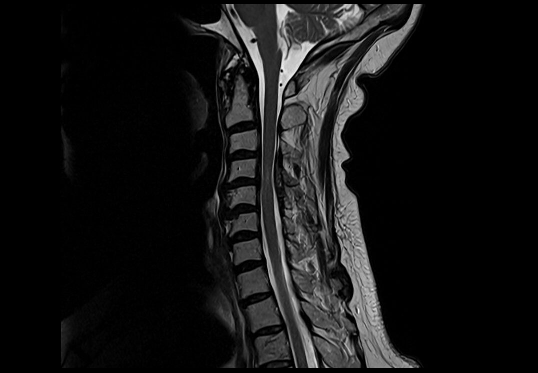
T2 sagittal sequence used in spine imaging
T2 axial sequence used in chest imaging

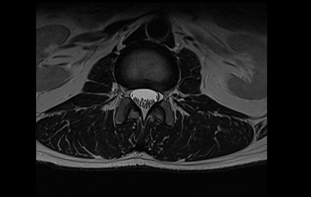
T2 axial sequence used in spine imaging
T2 axial sequence used in abdomen imaging

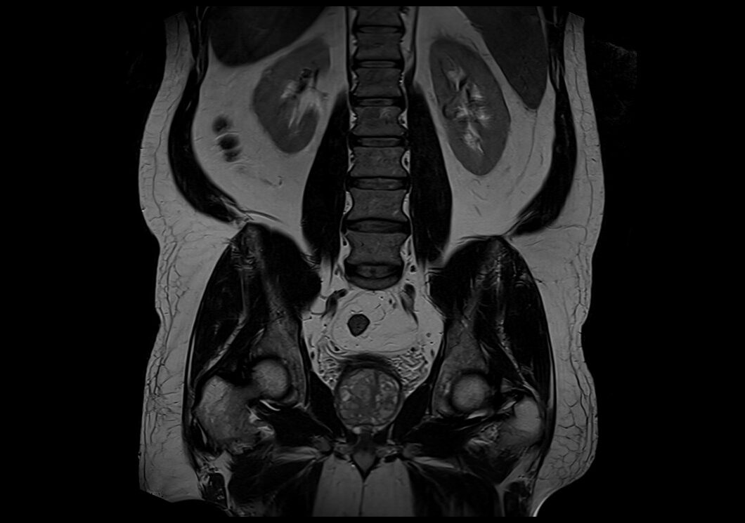
T2 coronal sequence used in abdominal imaging
T2 coronal sequence used in psoas imaging

T2 sagittal sequence used in L spine imaging

T2 axial sequence used in abdomen imaging

T2 axial sequence used in pelvic imaging

T2 sagittal sequence used in female pelvic imaging

T2 coronal sequence used in prostate imaging

T2 sagittal sequence used in hip imaging

T2 coronal sequence used in prostate imaging

T2 axial sequence used in knee imaging

T2 sagittal sequence used in upper arm imaging
