True Fisp (Fiesta / Balanced Sarge / Basg / True SSFP / STERF) FAT SAT
MRI image appearance
The easiest way to identify T2 TrueFISP fat-saturated images is to look for adipose tissues in the body, such as subcutaneous fat and fat in the bone marrow. Areas containing adipose tissues appear dark on T2 TrueFISP fat-saturated images. All other characteristics of the T2 TrueFISP fat-saturated images remain the same as those of the T2 TrueFISP images.
Tissues and their T2 TrueFISP fat saturated images appearance
Bone marrow: – dark
Muscles- gray
Fat – dark
White matter – dark gray
Moving blood- bright
Gray matter – gray
Fluids – bright
Bone – dark
Air – dark
Use
- Very Useful for pancreas imaging
- Very Useful for kidney imaging
- Very Useful for abdominal imaging
- Very useful for MRCP imaging
- Very useful for urography imaging
- Useful for chest imaging
- Useful for small bowel imaging
- Useful for pelvic imaging
Pathological appearance
Pathologies with adipose tissue content will appear dark on T2 TrueFISP fat-saturated images (e.g. lipoma). Due to the added fat suppression, pathological processes are usually very bright on T2 TrueFISP fat-saturated images.
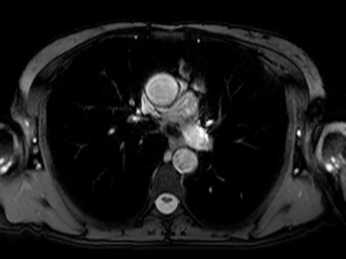
True FISP fat saturated axial sequence used in chest imaging
TRUE FISP FAT SATURATED AXIAL SEQUENCE USED IN UROGRAPHY IMAGING
