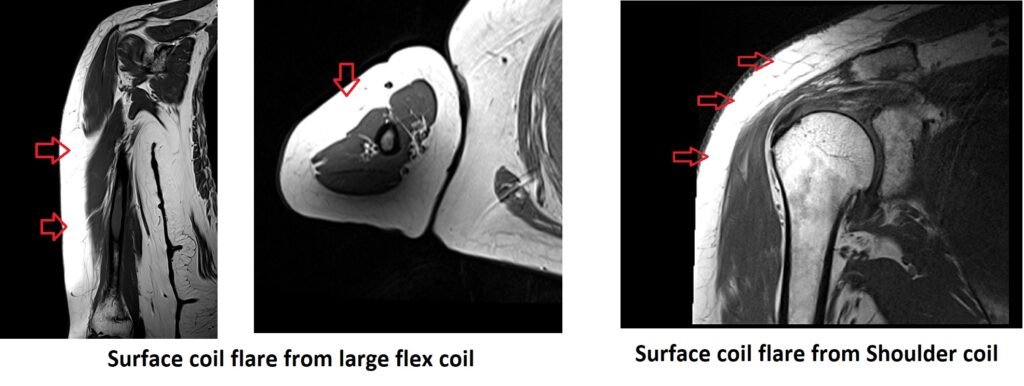
MRI Surface Coil Flare Artifact
The surface coil flare artifact is a common artifact that can occur in MRI imaging when using surface coils. It appears as a region of increased signal intensity surrounding the area directly under the surface coil. This artifact occurs due to the selective intensification of signals originating from superficial regions in the patient, while signals originating from deeper regions are simultaneously weakened or attenuated.
The surface coil flare artifact can have several implications:
Loss of image contrast: The flare artifact can lead to a loss of image contrast within the affected region, making it challenging to differentiate structures or abnormalities.
Misinterpretation of pathology: The increased signal intensity caused by the flare artifact can mimic or obscure underlying pathology, leading to potential misinterpretation of the MRI findings.
Limitations in evaluation: The presence of the flare artifact can hinder the accurate assessment of adjacent structures or areas of interest, reducing the diagnostic confidence of the MRI examination.
Here are some strategies to minimize or avoid The surface coil flare:
Repositioning the coil: Adjust the position and orientation of the surface coil to minimize its direct interaction with the region of interest. Experiment with different coil placements to find the configuration that produces the least amount of flare artifact.
Use of additional RF shielding: Applying additional RF shielding materials, such as foam or non-conductive fabric, between the coil and the patient’s body, can help reduce the extent of the flare artifact.
Gradient reversal technique: Some MRI systems offer a gradient reversal technique that can help minimize the surface coil flare artifact. By reversing the gradient polarity during the acquisition, the artifact’s location and intensity can be altered, potentially reducing its impact on the final image.
Use of alternative coils or sequences: Consider using different coil types, such as phased-array coils or volume coils, which may produce less pronounced flare artifacts. Additionally, alternative imaging sequences, such as spin echo or inversion recovery, may be less susceptible to the artifact compared to gradient echo sequences.
Post-processing techniques: If the flare artifact is still present in the acquired images, post-processing techniques, such as image filtering or correction algorithms, can be employed to reduce its visibility and improve image quality.
References:
- Ren H, Lin W, Ding X. Surface Coil Intensity Correction in Magnetic Resonance Imaging in Spinal Metastases. Med (Kaunas). 2017;12:138-143. doi: 10.1515/med-2017-0021. PMID: 28730173; PMCID: PMC5471916.
- Alves, F. C., & Berry, E. (2012). MRI Artifacts: Identification and Correction. Springer.