Musculoskeletal(MSK) Pelvis MRI Protocol and Planning
Indications for MRI MSK pelvis
- Identification and characterization of a wide variety of tumors affecting the bones, muscles, or soft tissues, including both benign and malignant tumors.
- Diagnosis and evaluation of infections involving joints, bones, and the surrounding soft tissues.
- Evaluation of muscles and tendons for injuries or abnormalities.
- Evaluation for bone infarction (avascular necrosis)
- Traumatic fracture
- Pathologic fracture
- Stress fracture
- Osteoarthritis
- Muscle injury
- Myositis
- Bursitis
- Cellulitis
Contraindications
- Any electrically, magnetically or mechanically activated implant (e.g. cardiac pacemaker, insulin pump biostimulator, neurostimulator, cochlear implant, and hearing aids)
- Intracranial aneurysm clips (unless made of titanium)
- Pregnancy (risk vs benefit ratio to be assessed)
- Ferromagnetic surgical clips or staples
- Metallic foreign body in the eye
- Metal shrapnel or bullet
Patient preparation for Musculoskeletal(MSK) Pelvis MRI
- A satisfactory written consent form must be taken from the patient before entering the scanner room
- Ask the patient to remove all metal object including keys, coins, wallet, any cards with magnetic strips, jewellery, hearing aid and hairpins
- Ask the patient to undress and change into a hospital gown
- If possible provide a chaperone for claustrophobic patients (e.g. relative or staff )
- Offer earplugs or headphones, possibly with music for extra comfort
- Explain the procedure to the patient
- Instruct the patient to keep still
- Note the weight of the patient
Positioning for Musculoskeletal(MSK) Pelvis MRI
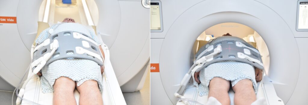
- Position the patient in supine position with head pointing towards the magnet (head first supine)
- Place the patient over the spine coil and position the body coil over the pelvis, aligning it from 2 inches above the iliac crest to 2 inches below the pubic symphysis
- Securely tighten the body coil using straps to prevent respiratory artefacts
- Give a pillow under the head for extra comfort (do not give cushions under the legs)
- Centre the laser beam localiser over hip joints (4 inches below iliac crest)
Recommended Musculoskeletal(MSK) Pelvis MRI Protocols and Planning
MSK Pelvis MRI Localiser
A three-plane T2 HASTE/T1 GRE localizer must be taken at the beginning to localize and plan the sequences. Typically, localizers take less than 25 seconds, and you have the option to use either T2 HASTE or T1 GRE low-resolution scans for this purpose.

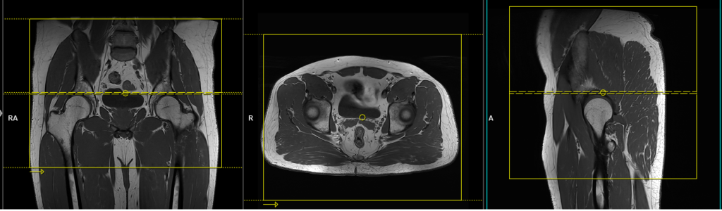
T1 tse coronal 4mm
Plan the coronal slices on the axial plane; angle the positioning block parallel to the right and left femoral heads. Check the positioning block in the other two planes. An appropriate angle must be given in the sagittal plane (vertically across the pelvis or parallel to the femur in a straight leg position). The slices should sufficiently cover the entire pelvis, extending from the anterior abdominal wall to the buttock region. The field of view (FOV) should be large enough to accommodate the bony pelvis, spanning from 2 inches above the iliac crest to two inches below the pubic symphysis.
Parameters
TR 500-700 | TE 15-25 | SLICE 4 MM | FLIP 150 | PHASE R>L | MATRIX 512X448 | FOV 350-380 | GAP 10% | NEX(AVRAGE) 2 |
T2 stir coronal 4mm
Plan the coronal slices on the axial plane; angle the positioning block parallel to the right and left femoral heads. Check the positioning block in the other two planes. An appropriate angle must be given in the sagittal plane (vertically across the pelvis or parallel to the femur in a straight leg position). The slices should sufficiently cover the entire pelvis, extending from the anterior abdominal wall to the buttock region. The field of view (FOV) should be large enough to accommodate the bony pelvis, spanning from 2 inches above the iliac crest to two inches below the pubic symphysis.
Parameters
TR 5000-6000 | TE 110 | FLIP 150 | NEX 2 | SLICE 4 MM | MATRIX 384X320 | FOV 350-380 | PHASE R>L | GAP 10% | TI 150 |
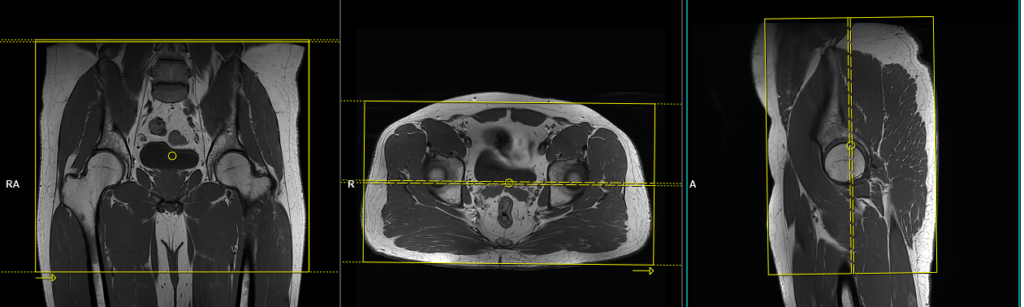
T1 tse axial 4mm
Plan the axial slices on the coronal plane and angle the positioning block parallel to both the right and left femoral heads. Check the positioning block in the other two planes to ensure an appropriate angle is maintained in the sagittal plane. This angle should be either horizontally across the pelvis or perpendicular to the femur in a straight leg position. The slices must be sufficient to cover the MSK pelvis from 1 inch above the iliac crest to 2 inches below the pubic symphysis. The FOV (field of view) must be large enough to accommodate the whole pelvis, usually ranging from 350 to 450mm.
Parameters
TR 600-700 | TE 15-25 | SLICE 4MM | FLIP 150 | PHASE R>L | MATRIX 512X448 | FOV 350-450 | GAP 10% | NEX(AVRAGE) 2 |
T2 stir axial 3mm
Plan the axial slices on the coronal plane and angle the positioning block parallel to both the right and left femoral heads. Check the positioning block in the other two planes to ensure an appropriate angle is maintained in the sagittal plane. This angle should be either horizontally across the pelvis or perpendicular to the femur in a straight leg position. The slices must be sufficient to cover the MSK pelvis from 1 inch above the iliac crest to 2 inches below the pubic symphysis. The FOV (field of view) must be large enough to accommodate the whole pelvis, usually ranging from 350 to 450mm.

Parameters
TR 6000-7000 | TE 110 | FLIP 150 | NEX 2 | SLICE 4 MM | MATRIX 384X320 | FOV 350-450 | PHASE R>L | GAP 10% | TI 150 |
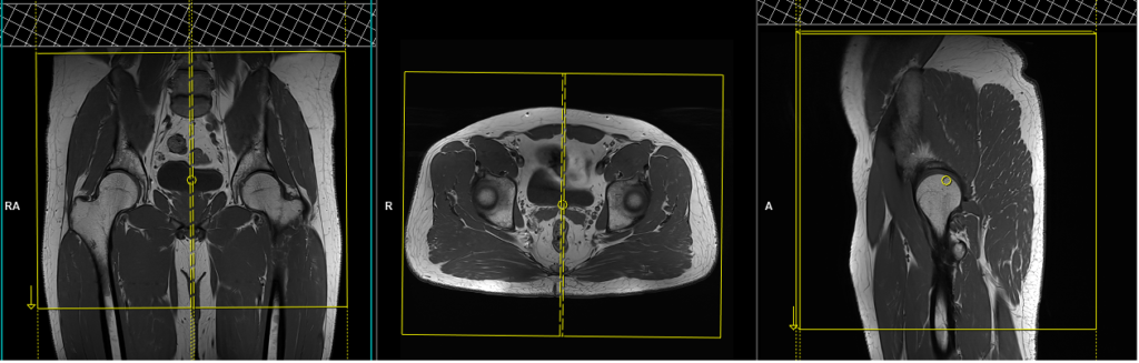
T2 tse sagittal 5mm
Plan the sagittal slices on the coronal plane; angle the positioning block parallel to the lumbar spine and pubic symphysis. Check the positioning block in the other two planes. An appropriate angle must be given in the axial plane (parallel to the linea alba and median sacral crest). Slices must be sufficient to cover the entire pelvis from right to left. The FOV (field of view) must be large enough to accommodate the whole pelvis, usually ranging from 350 to 450mm.
Parameters
TR 6000-7000 | TE 100-120 | SLICE 5 MM | FLIP 150-160 | PHASE H>F | MATRIX 515X512 | FOV 350-450 | GAP 10% | NEX(AVRAGE) 2 |