Contrast Media Related Artifacts
Contrast media artifacts in MRI are image artifacts that occur as a result of administering a contrast agent during a scan. These artifacts arise due to changes in the magnetic properties of tissues caused by the presence of the contrast agent, leading to alterations in the signal and distortions in the resulting images. Here are some common contrast media artifacts in MRI
Here are some common contrast media artifacts in MRI:
Susceptibility artifacts: Contrast agents can introduce susceptibility differences in tissues, causing local magnetic field distortions and resulting in signal loss or signal pile-up artifacts. These artifacts are more commonly observed near air-tissue interfaces or in regions with large susceptibility variations.
Image non-uniformity: Contrast agents can sometimes lead to image non-uniformity or shading artifacts, where the signal intensity varies across the image. This can be due to variations in contrast agent concentration or injection technique.
Chemical shift artifacts: Some contrast agents, particularly those containing fat components, can create chemical shift artifacts. These artifacts manifest as signal misregistration between fat and water, resulting in ghosting or displacement of structures. They are more noticeable in sequences sensitive to chemical shift differences, such as gradient-echo imaging.
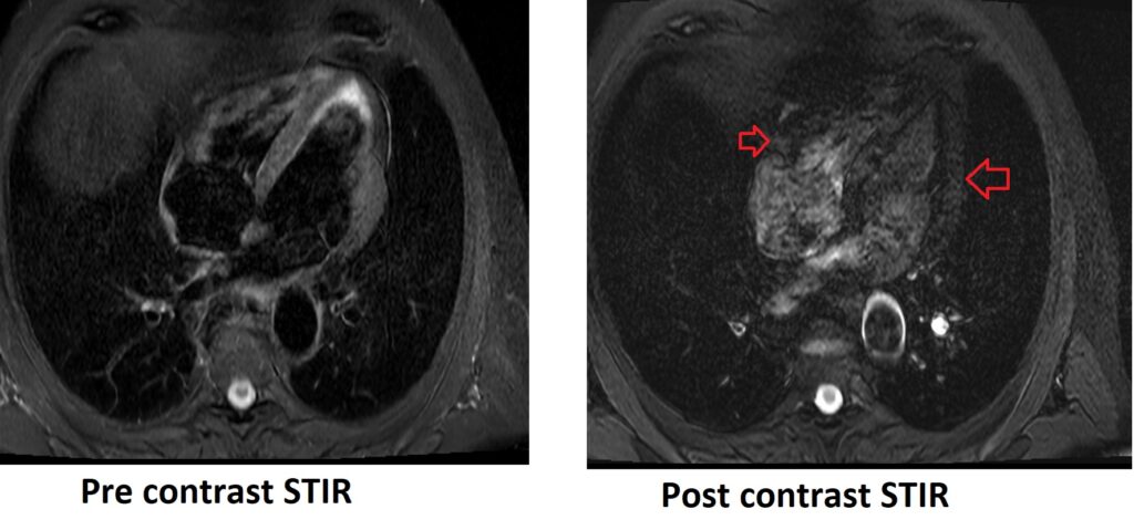
T1 shortening artifacts: Contrast agents with a short T1 relaxation time can cause T1 shortening artifacts. These artifacts can result in increased signal intensity in T1-weighted images, making it challenging to differentiate enhancing structures from surrounding tissues.
References:
- Ferreira, P. F., Gatehouse, P. D., Mohiaddin, R. H., & Firmin, D. N. (2013). Cardiovascular magnetic resonance artefacts. Journal of Cardiovascular Magnetic Resonance, 15, 41.
- Pettersson, H., Ackerman, N., Kaude, J., Googe, R. E., Mancuso, A. A., & Scott, K. N. (1986). Gadolinium-DTPA Enhancement of Experimental Soft Tissue Carcinoma and Hemorrhage in Magnetic Resonance Imaging. Magnetic Resonance Imaging, 4(1), 75-78.
- Mai, W. (2008). Pseudolayering Artefact on Postcontrast Magnetic Resonance Images of the Bladder of 18 Dogs and Three Cats. Vet Rec, 163(4), 117-119. doi:10.1136/vr.163.4.116.