MRI Thumb : Protocol and Planning
Indications for thumb MRI scan
- Marrow abnormalities (e.g. bone contusions, osteonecrosis, marrow oedema syndromes, and stress fractures)
- Synovial based disorders ( e.g. synovitis, tenosynovitis, bursitis, and ganglion cysts)
- Infections of bone, joint, or soft tissue (eg. osteomyelitis, osteo arthritis )
- Neoplasms of bone, joint or soft tissue
- Avascular necrosis
- Nerve Impingement
- Fractures
- Soft-tissue masses
- Occult fracture
- Ganglion cyst
- Ligament tear
Contraindications
- Any electrically, magnetically or mechanically activated implant (e.g. cardiac pacemaker, insulin pump biostimulator, neurostimulator, cochlear implant, and hearing aids)
- Intracranial aneurysm clips (unless made of titanium)
- Pregnancy (risk vs benefit ratio to be assessed)
- Ferromagnetic surgical clips or staples
- Metallic foreign body in the eye
- Metal shrapnel or bullet
Patient preparation for thumb MRI scan
- A satisfactory written consent form must be taken from the patient before entering the scanner room
- Ask the patient to remove all metal objects including keys, coins, wallet, cards with magnetic strips, jewellery, hearing aid and hairpins
- If possible provide a chaperone for claustrophobic patients (e.g. relative or staff )
- Offer earplugs or headphones, possibly with music for extra comfort
- Explain the procedure to the patient
- Instruct the patient to keep still
- Note the hight and weight of the patient
Positioning for thumb MRI scan
- Head first prone with arm up (superman position)
- Position the hand in the and and wrist coil or the large flex coil and immobilize it with cushions.
- Give cushions under the chest for extra comfort
- Centre the laser beam localiser over the metacarpophalangeal joint
- Register the patient o the scanner as 'head first supine'

Recommended Thumb MRI Protocols, Parameters, and Planning
Thumb MRI Localiser
A three-plane localizer must be taken at the beginning to localize and plan the sequences. Typically, these localizers take less than 25 seconds and can be achieved using T1 weighted low-resolution scans. It is advisable to obtain additional localizers until you have acquired accurate axial, coronal, and sagittal localizer images.

Localiser 2
Plan a three-plane thumb localizer on the coronal and axial localizer images. Plan the thumb coronal localizer on the hand coronal localizer. Align the planning block parallel to the metacarpal and phalanx bones. Verify the planning in the axial localizer and align the block parallel to the long axis of the metacarpal bones.
Next, plan the thumb sagittal localizer on the axial plane and orient it perpendicular to the long axis of the metacarpal bones. Confirm the planning in the sagittal hand localizer and align it parallel to the metacarpal and phalanx bones.
Finally, plan the axial thumb localizer on the coronal hand localizer and position it perpendicular to the long axis of the metacarpal bone. Verify the positioning on the sagittal hand localizer and align it perpendicular to the long axis of the metacarpal bone.

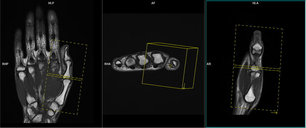
T2 stir axial 3mm SFOV(80mm)
Plan the axial slices on the sagittal thumb localizer and position the planning block perpendicular to the metacarpal and phalanx bones. Check the positioning block in the other two planes. Use an appropriate angle in the coronal localizer, ensuring it is perpendicular to the metacarpal and phalanx bones. The slices should be sufficient to cover the entire thumb, from the fingertips to the carpometacarpal joint of the thumb.
Parameters
TR 5000-6000 | TE 110 | FLIP 150 | NEX 3 | SLICE 3MM | MATRIX 224×224 | FOV 70-80 | PHASE A>P | GAP 10% | TI 160 |
Note:-An FOV ranging from 70mm to 80mm can only be achieved when utilizing a specialized high-channel wrist coil or conducting the scan within a 3T scanner. In cases where these choices are unavailable within your department, kindly opt for an FOV between 110mm and 130mm. The presented scans were executed using a 3T scanner equipped with deep resolution software.
T1 tse axial 3mm SFOV(80mm)
Plan the axial slices on the sagittal thumb localizer and position the planning block perpendicular to the metacarpal and phalanx bones. Check the positioning block in the other two planes. Use an appropriate angle in the coronal localizer, ensuring it is perpendicular to the metacarpal and phalanx bones. The slices should be sufficient to cover the entire thumb, from the fingertips to the carpometacarpal joint of the thumb.
Parameters
TR 400-600 | TE 15-25 | SLICE 3 MM | FLIP 150 | PHASE A>P | MATRIX 256X224 | FOV 70-80 | GAP 10% | NEX(AVRAGE) 3 |
T1 tse coronal 2mm SFOV(140X70mm)
Plan the coronal slices on the sagittal thumb localizer and angle the positioning block parallel to the metacarpal and phalanx bones. Check the positioning block in the other two planes. An appropriate angle must be used in the axial localizer, parallel to the long axis of metacarpal bones. The slices should be sufficient to cover the entire thumb, skin to skin. If available on your scanner, please use a smaller rectangular FOV of 70x140mm. In this case, use a phase direction from head to feet. If a rectangular FOV is not available, use a 140mm FOV with a right-to-left phase direction.
Parameters
TR 400-600 | TE 15-25 | SLICE 2 MM | FLIP 160 | PHASE H>F | MATRIX 256X256 | FOV 70×140 | GAP 10% | NEX(AVRAGE) 3 |
T2 stir coronal 2mm SFOV(140X70mm)
Plan the coronal slices on the sagittal thumb localizer and angle the positioning block parallel to the metacarpal and phalanx bones. Check the positioning block in the other two planes. An appropriate angle must be used in the axial images, parallel to the long axis of metacarpal bones. The slices should be sufficient to cover the entire thumb, skin to skin. If available on your scanner, please use a smaller rectangular FOV of 70x140mm. In this case, use a phase direction from head to feet. If a rectangular FOV is not available, use a 140mm FOV with a right-to-left phase direction.
Parameters
TR 3000-4000 | TE 110 | FLIP 150 | NEX 3 | SLICE 2 MM | MATRIX 256X224 | FOV 70×140 | PHASE H>F | GAP 10% | TI 160 |
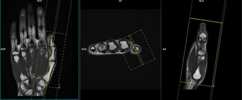
T2 stir sagittal 2mm SFOV(140X70mm)
Plan the sagittal slices on the coronal scans and angle the positioning block parallel to the metacarpal and phalanx bones. Check the positioning block in the other two planes. Ensure that an appropriate angle is used in the axial images, perpendicular to the long axis of the metacarpal bones. The slices should be sufficient to cover the entire thumb, from skin to skin. If a smaller rectangular FOV of 70x140mm is available on your scanner, please utilize it. In this case, use a phase direction from head to feet. If a rectangular FOV is not available, use a 140mm FOV with a right-to-left phase direction.

Parameters
TR 3000-4000 | TE 110 | FLIP 150 | NEX 3 | SLICE 2 MM | MATRIX 256X224 | FOV 70×140 | PHASE H>F | GAP 10% | TI 160 |