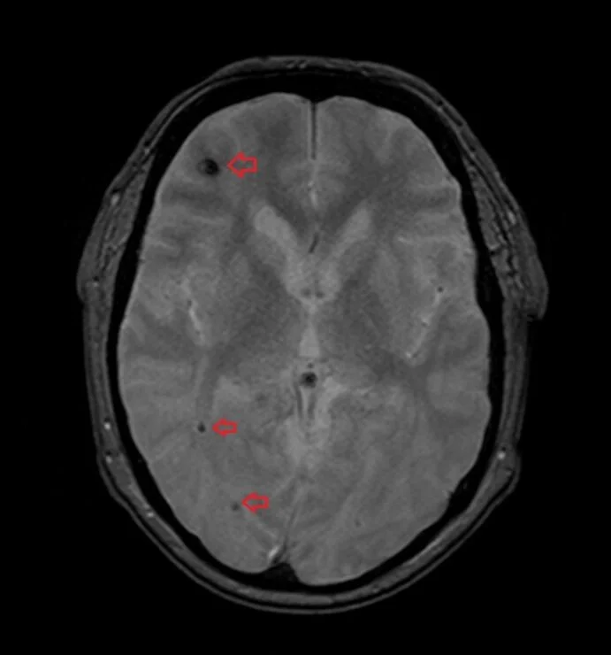
Blooming Artifact
Blooming artifact is a type of artifact that occurs in magnetic resonance imaging (MRI) when there are regions of high magnetic susceptibility differences. It appears as signal spreading or blooming beyond the actual boundaries of an object.

Blooming artifact in SWI:
Susceptibility Weighted Imaging (SWI) is a sequence that is particularly sensitive to magnetic susceptibility differences, making it useful for visualizing blood products, including hemorrhages. However, SWI can be susceptible to blooming artifact, which occurs when the signal from blood products spreads beyond their actual boundaries, leading to signal loss and distortion in surrounding structures.
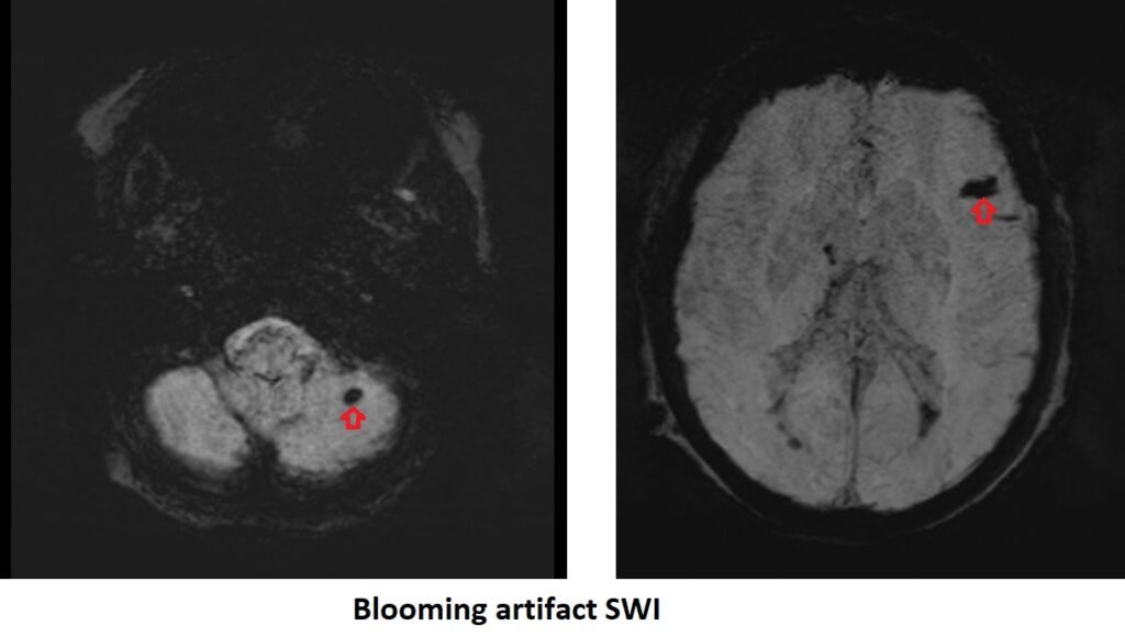

Blooming artifact SWI
Blooming artifact T2*(HEMO)
Here are some strategies to minimize or avoid Blooming Artifact
Sequence selection: Choosing the appropriate MRI sequence is crucial. Gradient-echo sequences, such as susceptibility-weighted imaging (SWI) or T2*-weighted sequences, are commonly used to detect hemorrhages due to their sensitivity to blood products. However, they are also more susceptible to blooming artifact. Spin-echo sequences, such as T1-weighted or T2-weighted sequences, may be preferred when blooming artifact is a concern.
Echo time optimization: Adjusting the echo time (TE) in gradient-echo sequences can help minimize blooming artifact. Shorter TE values can reduce blooming but may compromise sensitivity to certain types of hemorrhages. Longer TE values may improve visualization of hemorrhages but can increase the susceptibility to blooming. Finding the right balance is important.
Increase bandwidth: Increasing the receiver bandwidth can also help reduce the magnetic susceptibility artifact. A wider bandwidth allows for faster signal acquisition and better compensation for the magnetic field distortions.
References:
- Coward, J., Prier, J., Duda, J., & Yallapragada, A. (2020). Post‐thrombectomy intracranial blooming artifact. Clin Case Rep, 8(8), 1361–1364. doi: 10.1002/ccr3.2753.
- Saccaro, L. F., Bekri, I., De Malherbe, M., Hmida, I., & Pico, F. (2022). Ipsilateral blooming of microbleeds after Hyperintense Acute Reperfusion Marker sign in an ischemic Stroke patient: A case report. BMC Neurology, 22(142). Published on April 14, 2022.
- Nair, J.R., Van Hecke, W., De Belder, F., Venstermans, C., van den Hauwe, L., Van Goethem, J., & Parizel, P.M. (2010). High-Resolution Susceptibility-Weighted Imaging at 3 T With a 32-Channel Head Coil: Technique and Clinical Applications. Pictorial Essay. Neuroradiology/Head and Neck Imaging, 195(4)
- Weerink, L.B.M., Appelman, A.P.A., Kloet, R.W., & Van der Hoorn, A. (2022). Susceptibility-weighted imaging in intracranial hemorrhage: not all bleeds are black. The British Journal of Radiology, 96(1148).
- P. Holdsworth et al., “A review of MRI quality assurance: Needs, challenges, and solutions,” Journal of Magnetic Resonance Imaging, vol. 49, no. 2, pp. 374–383, 2019.