Wrist MRI Protocol and Planning
Indications for wrist MRI scan
- Marrow abnormalities (e.g. bone contusions, osteonecrosis, marrow oedema syndromes, and stress fractures)
- Synovial based disorders ( e.g. synovitis, tenosynovitis, bursitis, and ganglion cysts)
- Infections of bone, joint, or soft tissue (eg. osteomyelitis, osteo arthritis )
- Neoplasms of bone, joint or soft tissue
- Carpal tunnel syndrome
- Avascular necrosis
- Nerve Impingement
- Fractures in children
- Soft-tissue masses
- Carpal nonunions
- Occult fracture
- Ganglion cyst
- Ligament tear
Contraindications
- Any electrically, magnetically or mechanically activated implant (e.g. cardiac pacemaker, insulin pump biostimulator, neurostimulator, cochlear implant, and hearing aids)
- Intracranial aneurysm clips (unless made of titanium)
- Pregnancy (risk vs benefit ratio to be assessed)
- Ferromagnetic surgical clips or staples
- Metallic foreign body in the eye
- Metal shrapnel or bullet
Patient preparation for Wrist MRI
- A satisfactory written consent form must be taken from the patient before entering the scanner room
- Ask the patient to remove all metal objects including keys, coins, wallet, cards with magnetic strips, jewellery, hearing aid and hairpins
- If possible provide a chaperone for claustrophobic patients (e.g. relative or staff )
- Offer earplugs or headphones, possibly with music for extra comfort
- Explain the procedure to the patient
- Instruct the patient to keep still
- Note the weight of the patient
Positioning for Wrist MRI
- Head first prone with arm up (superman position)
- Position the wrist in the wrist coil or the small flex coil and immobilize it with cushions.
- Give cushions under the chest for extra comfort
- Centre the laser beam localiser over the wrist joint
- Register the patient in the scanner as head first supine

Recommended Wrist MRI Protocol Protocols and Planning
Localiser
A three-plane localizer must be taken at the beginning to localize and plan the sequences. Localizers are usually completed in less than 25 seconds. T1-weighted low-resolution scans are used for this purpose. A three-plane localizer must be taken at the beginning to localize and plan the sequences. Localizers are usually completed in less than 25 seconds. T1-weighted low-resolution scans are used for this purpose.

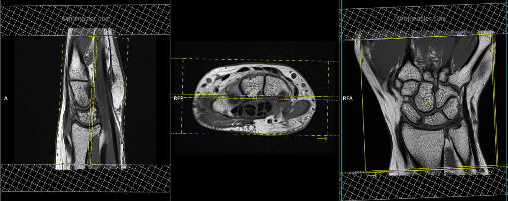
PD fat saturated axial 3mm SFOV
Plan the axial slices on the coronal plane; angle the positioning block horizontally across the wrist joint (i.e. across the carpal bones). Check the positioning block in the other two planes. An appropriate angle must be used in the sagittal plane (horizontally across the wrist joint). The slices should adequately cover the entire wrist area, from 1 inch above the carpo-metacarpal joint to 1 inch below the distal radioulnar joint. To mitigate vascular pulsation artifacts, employ saturation bands above and below the axial block.

Parameters
TR 3000-4000 | TE 15-25 | FLIP 150 | NEX 2 | SLICE 3 MM | MATRIX 288X288 | FOV 100-110 | PHASE R>L | GAP 10% | oversample 30% |
T2 stir coronal 3mm SFOV
Plan the coronal slices on the axial plane; angle the positioning block horizontally across the carpal bones. Verify the alignment of the positioning block in the other two planes. Utilize an adequate vertical angle in the sagittal plane to cover the wrist joint comprehensively from dorsal to palmar aspects. Additionally, implement saturation bands above and below the axial block to minimize vascular pulsation artifacts.
Parameters
TR 3000-4000 | TE 110 | FLIP 130 | NEX 2 | SLICE 3MM | MATRIX 256X256 | FOV 110-120 | PHASE H>F | GAP 10% | TI 130 |
T1 tse coronal 3mm SFOV
Plan the coronal slices on the axial plane; angle the positioning block horizontally across the carpal bones. Verify the alignment of the positioning block in the other two planes. Utilize an adequate vertical angle in the sagittal plane to cover the wrist joint comprehensively from dorsal to palmar aspects. Additionally, implement saturation bands above and below the axial block to minimize vascular pulsation artifacts.
Parameters
TR 400-600 | TE 15-25 | SLICE 3 MM | FLIP 130 | PHASE R>L | MATRIX 320X320 | FOV 110-120 | GAP 10% | NEX(AVRAGE) 2 |
PD fat saturated coronal 3mm SFOV
Plan the coronal slices on the axial plane; angle the positioning block horizontally across the carpal bones. Verify the alignment of the positioning block in the other two planes. Utilize an adequate vertical angle in the sagittal plane to cover the wrist joint comprehensively from dorsal to palmar aspects. Additionally, implement saturation bands above and below the axial block to minimize vascular pulsation artifacts.
Parameters
TR 3000-4000 | TE 15-25 | FLIP 150 | NEX 2 | SLICE 3 MM | MATRIX 288X288 | FOV 100-110 | PHASE R>L | GAP 10% | oversample 100% |
PD fat saturated sagittal 3mm SFOV
Plan the sagittal slices on the axial plane and angle the positioning block vertically across the carpal bones. Check the positioning block in the other two planes, ensuring an appropriate angle is used in the coronal plane, crossing the carpal bones vertically. The slices should sufficiently cover the entire wrist from medial to lateral aspects. The phase direction in the sagittal scans must be head to feet with 100% oversample to avoid artifacts from radial and ulnar artery pulsation. The use of saturation bands above and below the sagittal block will reduce radial and ulnar artery pulsation artifacts.

Parameters
TR 3000-4000 | TE 15-30 | FLIP 150 | NEX 2 | SLICE 3MM | MATRIX 288X288 | FOV 100-110 | PHASE H>F | GAP 10% | OVERSAMPLE 100% |