MRI Moiré Fringes Artifact
Moiré fringes are a common artifact observed in MRI images. This artifact typically occurs during gradient echo (GRE) sequences, especially when using body coils and when the patient is in contact with the edge of the magnet bore.
Moiré fringes arise due to a combination of factors, including the inhomogeneity of the main magnetic field and the wrap-around or aliasing of signals from areas outside the coil that contribute to the image formation. This leads to the superimposition and overlapping of images, resulting in the appearance of alternating light and dark bands along the edges of the image.
In terms of their visual appearance, Moiré fringes can be described as resembling “zebra stripes,” with the presence of distinct light and dark lines.

Here are some strategies to minimize or avoid Moiré Fringes Artifact
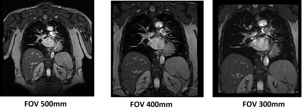
Small FOV: Moiré fringes often occur when the spatial frequency of the object being imaged matches the spatial frequency of the pixel grid in the MRI image. By using a smaller FOV, you can reduce the chances of this frequency matching and subsequently minimize the appearance of Moiré fringes.
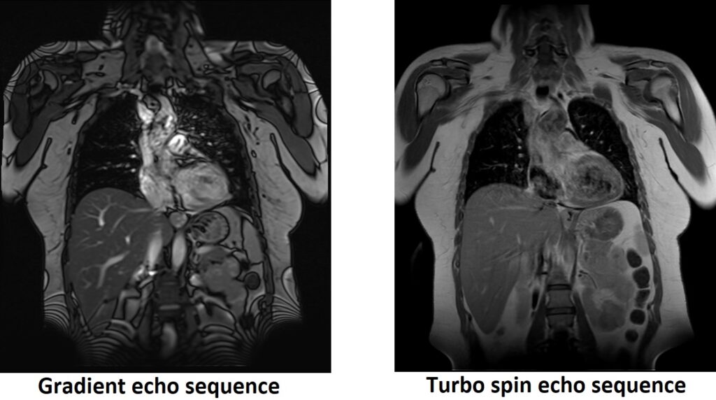
Alternative Sequences: Consider using alternative sequences such as turbo spin echo, which are less prone to this artifact.
References:
- Fink, J. R., Carr, R. B., & DeLong, D. M. (2013). Artifacts in body MR imaging: Their appearance and how to eliminate them. RadioGraphics, 33(2), 399-419.
- This reference provides an overview of various artifacts in body MRI, including Moiré fringes, and offers insights into their appearance and strategies for minimizing or eliminating them.
- Lauenstein, T. C., & Goehde, S. C. (2008). Body MRI: How to manage artifacts, motion, and metallic implants. European Radiology, 18(11), 1977-1987.