Cavernous Sinus MRI and MRA
Indications for cavernous sinus MRI
- Cavernous Sinus Thrombosis
- Cranial Nerve Dysfunction
- Tumors and Lesions
cavernous sinus anatomy
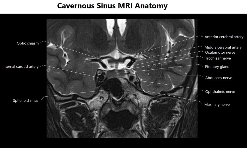
The cavernous sinus is a paired, venous structure located on each side of the sella turcica, a bony depression in the skull base. It plays a crucial role in the anatomy of the head and is involved in various critical functions.
Location: The cavernous sinus is situated on either side of the sella turcica, a saddle-shaped depression within the sphenoid bone at the base of the skull. It is centrally located within the cranial cavity.
Shape and Structure: The cavernous sinus is a complex, irregularly shaped structure. It is composed of a network of interconnected venous channels and sinuses, making it appear cavernous, hence the name.
Contents: The cavernous sinus houses several important structures, including:
Internal carotid artery: The internal carotid artery passes through the cavernous sinus as it travels toward the brain. The sinus helps protect and support this crucial blood vessel.
Cranial nerves: Several cranial nerves pass through or near the cavernous sinus, including cranial nerves III (oculomotor), IV (trochlear), V1 (ophthalmic division of the trigeminal nerve), V2 (maxillary division of the trigeminal nerve), and VI (abducens). These nerves control eye and facial movements, sensations, and autonomic functions.
Venous blood: The cavernous sinus receives venous blood from various sources, including the superior ophthalmic vein, the superficial middle cerebral vein, and the sphenoparietal sinus.
Function: The cavernous sinus serves several vital functions:
Blood drainage: It acts as a crucial pathway for draining blood from the brain, face, and eyes into the internal jugular vein, ultimately returning it to the heart.
Carotid artery protection: It cushions and protects the internal carotid artery, a major blood vessel supplying the brain.
Neurological control: The cavernous sinus plays a role in the functioning of cranial nerves, particularly those related to eye movements and facial sensations.
Clinical Significance: Lesions or abnormalities in the cavernous sinus can have significant clinical implications. Conditions such as cavernous sinus thrombosis (a blood clot within the sinus), tumors, or infections can affect the nerves passing through it, leading to symptoms such as vision changes, eye movement disorders, and facial pain.

Contraindications
- Any electrically, magnetically or mechanically activated implant (e.g. cardiac pacemaker, insulin pump biostimulator, neurostimulator, cochlear implant, and hearing aids)
- Intracranial aneurysm clips (unless made of titanium)
- Pregnancy (risk vs benefit ratio to be assessed)
- Ferromagnetic surgical clips or staples
- Metallic foreign body in the eye
- Metal shrapnel or bullet
Patient preparation for cavernous sinus MRI
- A satisfactory written consent form must be taken from the patient before entering the scanner room
- Ask the patient to remove all metal objects including keys, coins, wallet, cards with magnetic strips, jewellery, hearing aid and hairpins
- If possible provide a chaperone for claustrophobic patients (e.g. relative or staff )
- An intravenous line must be placed with extension tubing extending out of the magnetic bore
- Contrast injection risk and benefits must be explained to the patient before the scan
- Gadolinium should only be given to the patient if GFR is > 30
- Offer earplugs or headphones, possibly with music for extra comfort
- Explain the procedure to the patient
- Instruct the patient to keep still
- Note the height and weight of the patient
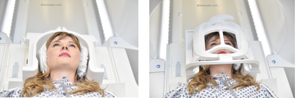
Positioning for cavernous sinus MRI
- Head first supine
- Position the head in the head coil and immobilise with cushions
- Give cushions under the legs for extra comfort
- Centre the laser beam localizer over the glabella
Recommended Cavernous Sinus Protocols and Planning
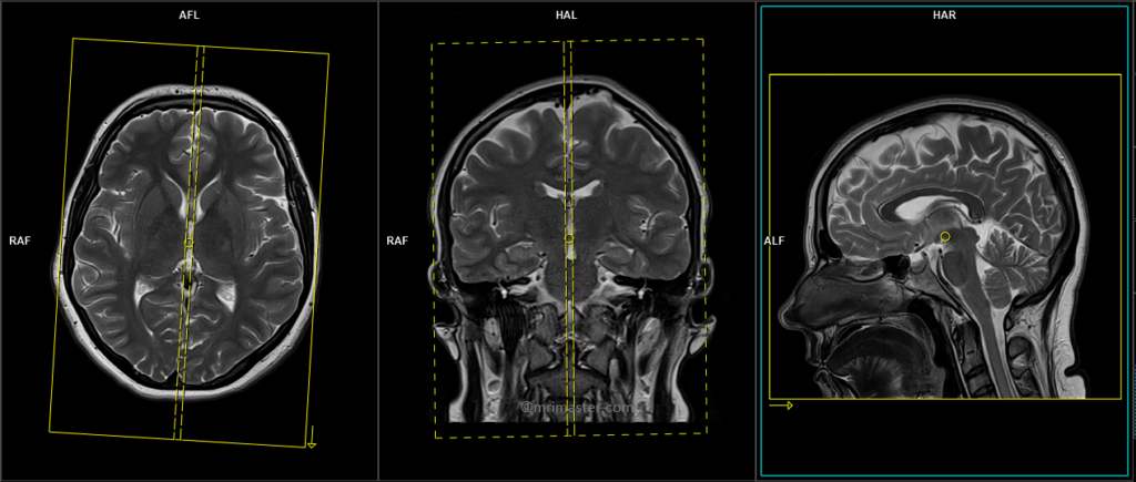
localiser
A three-plane localizer must be taken at the beginning to localize and plan the sequences. Localizers are usually less than 25 seconds and are T1-weighted low-resolution scans.

T2 tse axial 3mm
Plan the axial slices on the sagittal plane and position the block parallel to the genu and splenium of the corpus callosum. Verify the planning block in the other two planes and ensure that an appropriate angle is maintained in the coronal plane, making it perpendicular to the line of the midline of the brain and the 4th ventricle. Ensure that the number of slices is sufficient to cover the entire brain from the vertex to the line of the foramen magnum.

Parameters
TR 5000-6000 | TE 100-120 | SLICE 3MM | FLIP 130-150 | PHASE R>L | MATRIX 320X288 | FOV 210-230 | GAP 10% | NEX(AVRAGE) 2 |
T2 tse sagittal 3mm
Plan the sagittal slices on the axial plane and position the block parallel to the midline of the brain. Verify the planning block in the other two planes. Angle the planning block appropriately in the coronal plane, ensuring it is parallel to the line along the midline of the brain and the 4th ventricle. Make sure that the number of slices is sufficient to cover the entire brain from one temporal lobe to the other.
Parameters
TR 4500-6000 | TE 100-120 | SLICE 3MM | FLIP 130-150 | PHASE A>P | MATRIX 320X304 | FOV 210-230 | GAP 10% | NEX(AVRAGE) 2 |
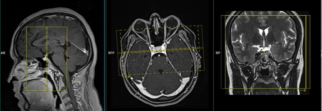
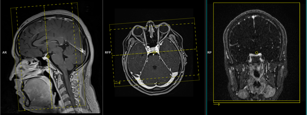
T2 tse 2mm coronal SFOV
Plan the coronal slices on the sagittal plane. Angle the planning block perpendicular to the sella turcica. Check the planning block in the other two planes. Ensure an appropriate angle is applied in the axial plane, which should be perpendicular to the midline of the brain. The slices must sufficiently cover the area of the cavernous sinus, extending from the mid ethmoid sinus to the fourth ventricle.
Parameters
TR 4000-5000 | TE 100-120 | SLICE 2 MM | FLIP 130-150 | PHASE R>L | MATRIX 256X256 | FOV 150-170 | GAP 10% | NEX(AVRAGE) 2 |
T1 VIBE DIXON axial .7mm pre contrast
Plan the axial slices on the sagittal plane and position the block parallel to the genu and splenium of the corpus callosum. Verify the planning of the block in the other two planes and ensure that an appropriate angle is maintained in the coronal plane, making it perpendicular to the midline of the brain. Make sure that the number of slices is sufficient to cover the cavernous sinus area from the corpus callosum to the hard palate.

Parameters
TR 6-7 | TE 2.39 4.77 | SLICE .7 MM | FLIP 12 | PHASE R>L | MATRIX 256X256 | FOV 270-290 | GAP 10% | NEX(AVRAGE) 2 |
TWIST coronal 0.9mm 3pre and 27 post
Plan the coronal slices along the sagittal plane. Angle the planning block perpendicular to the sella turcica. Check the positioning in the other two planes. Ensure an appropriate angle is applied in the axial plane, which should be perpendicular to the midline of the brain. The slices must sufficiently cover the area of the cavernous sinus, extending from the glabella to the fourth ventricle.
Parameters
TR 2-3 | TE 1.02 | FLIP 14 | NEX 1 | SLICE .9MM | MATRIX 204X204 | FOV 210-230 | PHASE R>L | DYNAMIC 30 SCAN | IPAT ON |
Twist MRA (Time-Resolved Angiography With Interleaved Stochastic Trajectories) is a dynamic imaging technique for real-time visualization of blood vessels . A dynamic TWIST sequence consists of 30 0.8mm 3D scans, with each acquisition taking around 3-4 seconds. The contrast injection must be administered after the third dynamic sequence.
TWIST MRA MIP images

T1 VIBE DIXON axial .7mm post contrast
Plan the axial slices on the sagittal plane and position the block parallel to the genu and splenium of the corpus callosum. Verify the planning of the block in the other two planes and ensure that an appropriate angle is maintained in the coronal plane, making it perpendicular to the midline of the brain. Make sure that the number of slices is sufficient to cover the cavernous sinus area from the corpus callosum to the hard palate.
Parameters
TR 6-7 | TE 2.39 4.77 | SLICE .7 MM | FLIP 12 | PHASE R>L | MATRIX 256X256 | FOV 270-290 | GAP 10% | NEX(AVRAGE) 2 |
VIBE DIXON axial post contrast images of cavernous sinus
