MRI Hip (unilateral)
Indications for MRI hips
- Intra or extra articular abnormality (e.g., loose body)
- Slipped femoral capital epipysis
- Tears of the acetabular labrum.
- Evaluate integrity of hip cartilage
- Degenerative disk disease
- Avascular necrosis
- Inflammatory arthritis
- Traumatic fracture
- Pathologic fracture
- Stress fracture
- Osteoarthritis
- Muscle injury
- Tendonitis
- Myositis
- Bursitis
- Cellulitis
Contraindications
- Any electrically, magnetically or mechanically activated implant (e.g. cardiac pacemaker, insulin pump biostimulator, neurostimulator, cochlear implant, and hearing aids)
- Intracranial aneurysm clips (unless made of titanium)
- Pregnancy (risk vs benefit ratio to be assessed)
- Ferromagnetic surgical clips or staples
- Metallic foreign body in the eye
- Metal shrapnel or bullet
Patient preparation for Hip MRI
- A satisfactory written consent form must be taken from the patient before entering the scanner room
- Ask the patient to remove all metal object including keys, coins, wallet, any cards with magnetic strips, jewellery, hearing aid and hairpins
- Ask the patient to undress and change into a hospital gown
- If possible provide a chaperone for claustrophobic patients (e.g. relative or staff )
- Offer earplugs or headphones, possibly with music for extra comfort
- Explain the procedure to the patient
- Instruct the patient to keep still
- Note the weight of the patient
Positioning for Hip MRI
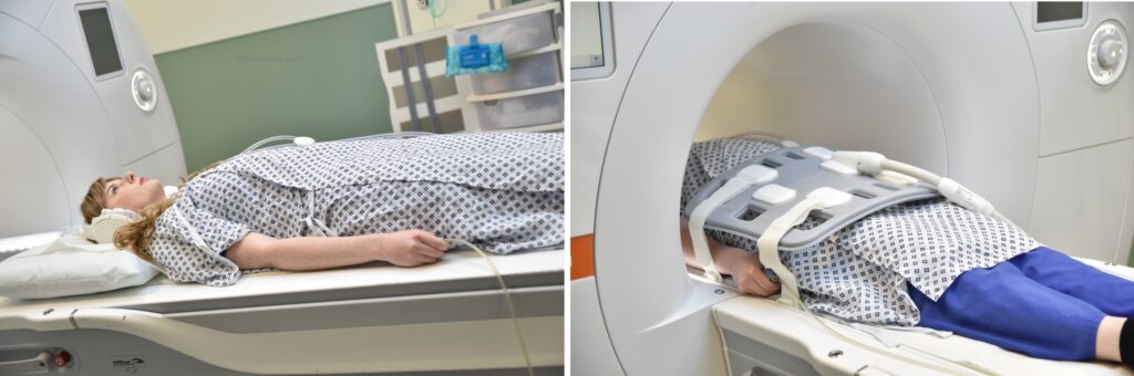
- Position the patient in supine position with head pointing towards the magnet (head first supine)
- Position the patient over the spine coil and place the body coil over the pelvis (iliac crest down to mid thigh)
- Securely tighten the body coil using straps to prevent respiratory artefacts
- Give a pillow under the head for extra comfort (do not give cushions under the legs)
- Centre the laser beam localiser over hip joints (4 inches below iliac crest)
Recommended Hip MRI Protocols, Parameters, and Planning
Hip MRI localiser
A three-plane localizer must be taken at the beginning to localize and plan the sequences. Localizers are normally less than 25 seconds, T1-weighted low-resolution scans.

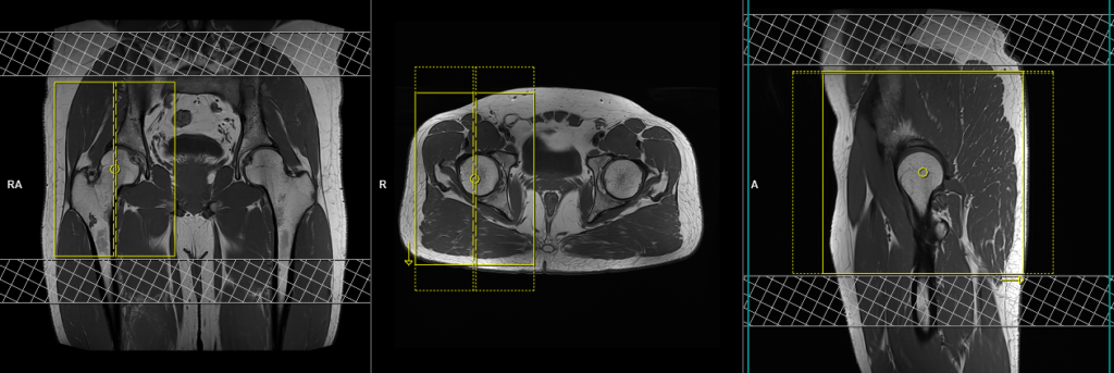
T1 tse coronal 3mm SFOV unilateral
Plan the coronal slices on the axial plane. Angle the positioning block perpendicular to the acetabulum (i.e., parallel to the femoral head and neck). Check the positioning block in the other two planes. An appropriate angle must be given in the sagittal plane (perpendicular to the acetabulum). The slices should adequately encompass the hip joint, starting from the ischial tuberosities and extending up to the line of the pubic symphysis. To minimize ghosting artifacts resulting from peristalsis, vascular pulsation, and breathing, consider incorporating a saturation band at the top and bottom of the coronal block.

Parameters
TR 400-600 | TE 15-25 | SLICE 3 MM | FLIP 150 | PHASE R>L | MATRIX 320X320 | FOV 200-230 | GAP 10% | NEX(AVRAGE) 3 |
T2 stir\PD fat saturated coronal 3mm SFOV unilateral
Plan the coronal slices on the axial plane. Angle the positioning block perpendicular to the acetabulum (i.e., parallel to the femoral head and neck). Check the positioning block in the other two planes. An appropriate angle must be given in the sagittal plane (perpendicular to the acetabulum). The slices should adequately encompass the hip joint, starting from the ischial tuberosities and extending up to the line of the pubic symphysis. To minimize ghosting artifacts resulting from peristalsis, vascular pulsation, and breathing, consider incorporating a saturation band at the top and bottom of the coronal block.
Parameters
TR 4000-5000 | TE 110 | FLIP 150 | NEX 2 | SLICE 3 MM | MATRIX 256X240 | FOV 200-230 | PHASE R>L | GAP 10% | TI 150 |
T1 tse axial 3mm SFOV unilateral
Plan the axial slices on the coronal plane and angle the positioning block horizontally across the femoral heads. Check the positioning block in the other two planes to ensure an appropriate angle is maintained in the sagittal plane, which should be perpendicular to the femur. The slices must be sufficient to cover the hip joints from 1 inch above the anterior inferior iliac spine to 1 inch below the lesser trochanter. To minimize ghosting artifacts resulting from peristalsis, vascular pulsation, and breathing, consider incorporating a saturation band at the top and bottom of the axial block.

Parameters
TR 400-600 | TE 15-25 | SLICE 3 MM | FLIP 160 | PHASE A>P | MATRIX 320X320 | FOV 200-230 | GAP 10% | NEX(AVRAGE) 3 |
T2 stir\PD fat saturated axial 3mm SFOV unilateral
Plan the axial slices on the coronal plane and angle the positioning block horizontally across the femoral heads. Check the positioning block in the other two planes to ensure an appropriate angle is maintained in the sagittal plane, which should be perpendicular to the femur. The slices must be sufficient to cover the hip joints from 1 inch above the anterior inferior iliac spine to 1 inch below the lesser trochanter. To minimize ghosting artifacts resulting from peristalsis, vascular pulsation, and breathing, consider incorporating a saturation band at the top and bottom of the axial block.
Parameters
TR 4000-5000 | TE 110 | FLIP 150 | NEX 2 | SLICE 3 MM | MATRIX 256X240 | FOV 200-230 | PHASE A>P | GAP 10% | TI 150 |
T2 TSE sagittal 3mm SFOV unilateral
Plan the sagittal slices on the coronal plane; angle the position block parallel to the femur. Check the positioning block in the other two planes. An appropriate angle must be given in the axial plane (perpendicular to the femoral head). Slices must be sufficient to cover the hip joint from outer cortex of the greater trochanter up to the inner portion of the acetabulum. To minimize ghosting artifacts caused by peristalsis and breathing, consider using a saturation band over the axial block.
Parameters
TR 3000-4000 | TE 100-120 | SLICE 3 MM | FLIP 130-150 | PHASE A>P | MATRIX 320X320 | FOV 200-230 | GAP 10% | NEX(AVRAGE) 2 |
Optional Scans
PD fat sat axial oblique 3mm SFOV affected side
Plan the axial oblique slices on the coronal plane, angling the positioning block parallel to the femoral neck. Verify the positioning block alignment in the other two planes. Ensure an appropriate angle is established in the axial plane, perpendicular to the femoral head. The slices should adequately cover the hip joint, spanning one inch above the superior border of the acetabulum to one inch below the inferior border of the acetabulum. To minimize ghosting artifacts caused by peristalsis and breathing, consider using a saturation band over the axial oblique block.
Parameters
TR 3000-4000 | TE 15-20 | SLICE 3 MM | FAT SAT ON | PHASE A>P | MATRIX 256X256 | FOV 200-250 | GAP 10% | NEX(AVRAGE) 2 |
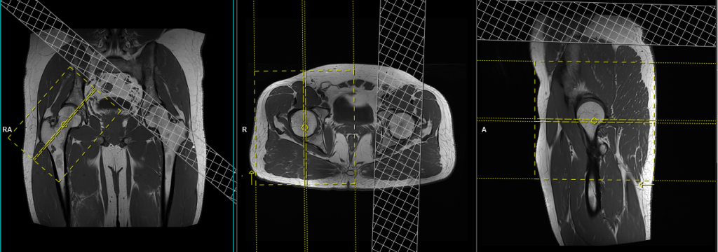
PD fat sat sagittal oblique 3mm SFOV affected side
Plan the sagittal oblique slices on the coronal plane; angle the positioning block perpendicular to the femoral neck. Check the positioning block in the other two planes. Ensure an appropriate angle is set in the axial plane (perpendicular to the femoral head). The slices should sufficiently cover the hip joint, extending one inch above the acetabulum to one inch below the greater trochanter. To minimize ghosting artifacts caused by peristalsis and breathing, consider using a saturation band over the sagittal oblique block.

Parameters
TR 3000-4000 | TE 15-20 | SLICE 3 MM | FAT SAT ON | PHASE A>P | MATRIX 256X256 | FOV 200-250 | GAP 10% | NEX(AVRAGE) 2 |
PD fat saturated SPACE 3D axial 1mm ISO SFOV unilateral
Plan the axial 3D block on the coronal plane and angle the positioning block horizontally across the femoral heads. Check the positioning block in the other two planes to ensure an appropriate angle is maintained in the sagittal plane, which should be perpendicular to the femur. The slices must be sufficient to cover the hip joints from 1 inch above the anterior inferior iliac spine to 1 inch below the lesser trochanter. To minimize ghosting artifacts resulting from peristalsis, vascular pulsation, and breathing, consider incorporating a saturation band at the top and bottom of the axial block.
