Fetal Brain MRI
Indications for fetal brain MRI scan
- ABDOMINAL WALL DEFECTS AND TUMORS
- Management of arachnoid cysts
- Sacrococcygeal teratomas
- Processes obstructing the airway (such as neck mass or congenital high airway obstruction)
- Complications of monochorionic twinning
- Chest masses
Contraindications
- Any electrically, magnetically or mechanically activated implant (e.g. cardiac pacemaker, insulin pump biostimulator, neurostimulator, cochlear implant, and hearing aids)
- Intracranial aneurysm clips (unless made of titanium)
- Ferromagnetic surgical clips or staples
- Metallic foreign body in the eye
- Metal shrapnel or bullet
Patient preparation for fetal brain MRI scan
- A satisfactory written consent form must be taken from the patient before entering the scanner room
- Ask the patient to remove all metal object including keys, coins, wallet, any cards with magnetic strips, jewellery, hearing aid and hairpins
- Ask the patient to undress and change into a hospital gown
- An intravenous line must be placed with extension tubing extending out of the magnetic bore
- Claustrophobic patients may be accompanied into the scanner room e.g. by staff member or relative with proper safety screening
- Offer earplug or headphones possibly with music for extra comfort
- Explain the procedure to the patient and answer questions
- Note down the hight and weight of the patient
- Pregnancy scanning consent must be taken before the procedure
Positioning for fetal brain MRI scan
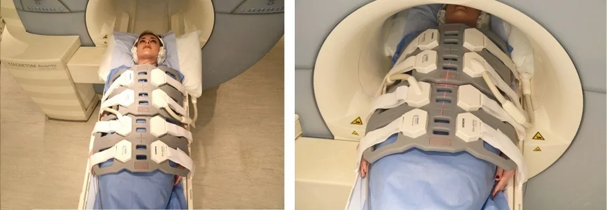
- Position the patient in supine position with head pointing towards the magnet (head first supine)
- Position the patient over the spine coil and place the body coils over abdomen and pelvis (nipple down to elbow three inches below symphysis pubis)
- Securely tighten the body coil using straps to prevent respiratory artefacts
- Give a pillow under the head and cushions under the legs for extra comfort
- Centre the laser beam localiser over mid abdomen
- Register the patient in the scanner as head first supine
Recommended MRI Fetal Brain Scan Protocols and Planning
Fetal Brain Scan localiser
A three-plane HASTE (Half-Fourier Acquisition Single-shot Turbo Spin Echo) localizer is commonly used in magnetic resonance imaging (MRI) to localize and plan sequences. HASTE localizers are fast and can be acquired in under 25 seconds, making them useful for localizing abdominal and pelvic structures.
To obtain the best results, it is generally recommended to acquire at least 3-4 slices in all planes (axial, sagittal, and coronal) during the localizer sequence. These slices help in accurately identifying the position and orientation of the anatomy of interest, which is important for planning subsequent imaging sequences.

T2 HASTE sagittal 6 mm Respiratory gated
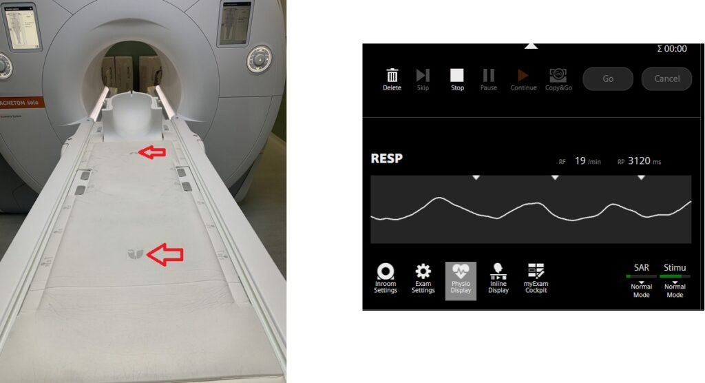
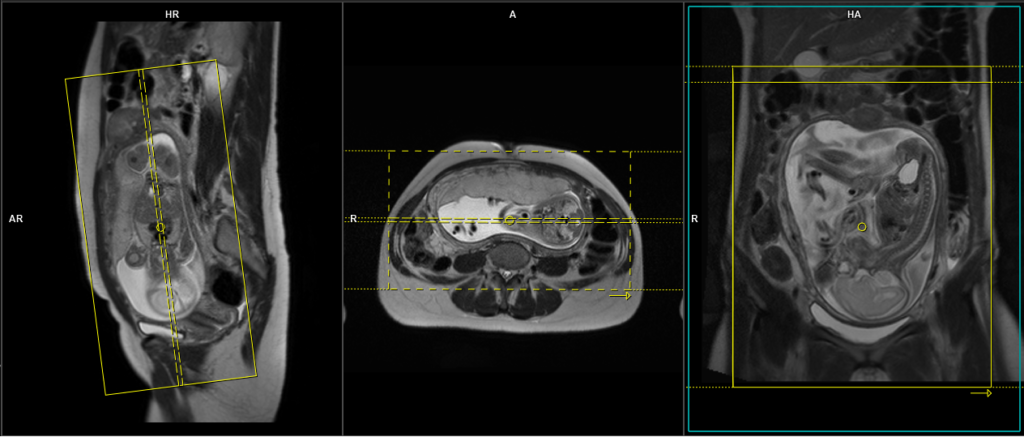
Begin by planning the sagittal slices on the coronal localizer and position the block parallel to the gravid uterus. Verify the positioning block in the other two planes to confirm proper alignment. It is essential to provide an appropriate angle in the axial plane, which should be perpendicular to the gravid uterus. The number of slices should be sufficient to cover the entire abdomen and pelvis, from right to left. The field of view (FOV) must be wide enough to encompass the whole abdomen and pelvis, typically ranging from 400 mm to 480 mm. However, it is important to note that these scans usually take approximately 35 to 40 seconds, which can be challenging for a pregnant woman to hold her breath. To address this issue, we perform the scan under respiratory gating. There are two options for respiratory gating: the liver dome method or the table respiratory sensors. In our department, we utilise the table respiratory sensor.s

Parameters
TR 4-5 | TE 2-3 | FLIP 60 | NEX 1 | SLICE 6 MM | MATRIX 320×320 | FOV 400-480 | PHASE R>L | OVERSAMPLE 30% | IPAT ON |
Table sensors
Advanced MRI scanners are equipped with built-in table sensors that detect the respiratory waveform and trigger data acquisition during the expiration phase of the respiratory cycle. Proper patient positioning over the sensor is critical for accurate respiratory gating. This method eliminates the need for external respiratory gating equipment, such as sensors and belts.
T2 HASTE coronal 6 mm Respiratory gated
Plan the coronal slices on the sagittal localizer and position the block parallel to the gravid uterus. Verify the positioning block in the other two planes for proper alignment. An appropriate angle should be set in the axial plane, running parallel across the gravid uterus. The number of slices should be sufficient to cover the entire abdomen and pelvis, from the anterior abdominal wall to the spinous process of the vertebrae. The field of view (FOV) must be large enough to encompass the entire abdomen and pelvis, typically ranging from 400 mm to 480 mm. However, it is important to note that these scans usually take approximately 30 to 35 seconds, which can be challenging for a pregnant woman to hold her breath. To address this issue, we perform the scan under respiratory gating. There are two options for respiratory gating: the liver dome method or the table respiratory sensors. In our department, we utilise the table respiratory sensor.
Parameters
TR 4-5 | TE 2-3 | FLIP 60 | NEX 1 | SLICE 6MM | MATRIX 320×320 | FOV 400-480 | PHASE R>L | OVERSAMPLE 30% | IPAT ON |
T2 HASTE axial 6 mm Respiratory gated
Plan the axial slices on the sagittal scans and angle the position block perpendicular through the gravid uterus. Verify the positioning block in the other two planes for proper alignment. An appropriate angle should be set in the coronal plane, running perpendicular across the gravid uterus. The number of slices should be sufficient to cover the entire abdomen and pelvis, from the diaphragm to the pubic symphysis. The field of view (FOV) must be large enough to encompass the entire abdomen and pelvis, typically ranging from 400 mm to 480 mm. However, it is important to note that these scans usually take approximately 40 to 45 seconds, which can be challenging for a pregnant woman to hold her breath. To address this issue, we perform the scan under respiratory gating. There are two options for respiratory gating: the liver dome method or the table respiratory sensors. In our department, we utilise the table respiratory sensor.
Parameters
TR 4-5 | TE 2-3 | FLIP 60 | NEX 1 | SLICE 5 MM | MATRIX 320×320 | FOV 400-480 | PHASE A>P | OVERSAMPLE 10% | IPAT ON |
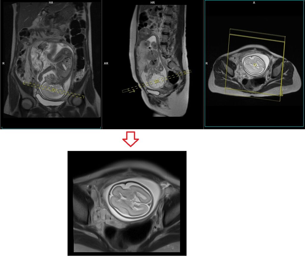
T2 HASTEcoronal localiser fetal BRAIN
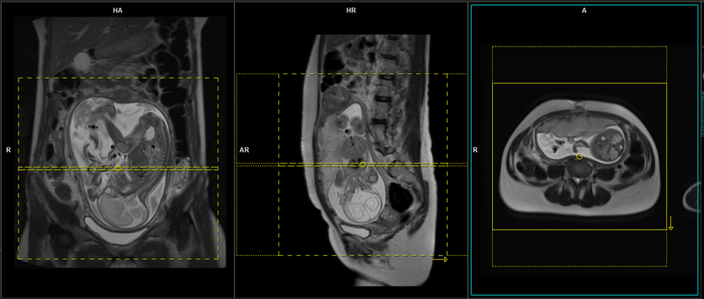
Plan the single-slice axial localizer on the coronal uterus scans. Angle the position block parallel to the genu and splenium of the corpus callosum of the fetal head. Verify the positioning block in the other two planes to ensure proper alignment. In the sagittal scans, establish an appropriate angle running perpendicular to the midline of the fetal brain. The field of view (FOV) must be sufficiently large to cover the entire brain, typically ranging from 180 mm to 250 mm. It is important to include an appropriate amount of oversampling to prevent any wrap-around artifacts. These scans usually take approximately 1 to 2 seconds, and they can be performed either as a breath-hold or using the respiratory sensor.
Please note that the position of the fetal head may vary among different patients and during different trimesters. Therefore, it is crucial to plan the localiser according to the position of the fetal brain. The reference points mentioned in the planning process remain the same.
Parameters
TR 4-5 | TE 2-3 | FLIP 60 | NEX 1 | SLICE 5 MM | MATRIX 320×320 | FOV 400-480 | PHASE A>P | OVERSAMPLE 10% | IPAT ON |
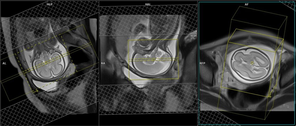
T2 HASTE sagittal localiser fetal brain
Plan the single-slice sagittal localizer on the axial uterus scans or axial localizer. Angle the position block parallel to the midline of the fetal head. Verify the positioning block in the other two planes to ensure proper alignment. In the coronal scans, establish an appropriate angle running parallel to the midline of the fetal brain. The field of view (FOV) must be sufficiently large to cover the entire brain, typically ranging from 180 mm to 250 mm. It is important to include an appropriate amount of oversampling to prevent any wrap-around artifacts. These scans usually take approximately 1 to 2 seconds, and they can be performed either as a breath-hold or using the respiratory sensor.
Please note that the position of the fetal head may vary among different patients and during different trimesters. Therefore, it is crucial to plan the localiser according to the position of the fetal brain. The reference points mentioned in the planning process remain the same.

Parameters
TR 4-5 | TE 2-3 | FLIP 60 | NEX 1 | SLICE 5 MM | MATRIX 320×320 | FOV 400-480 | PHASE A>P | OVERSAMPLE 10% | IPAT ON |
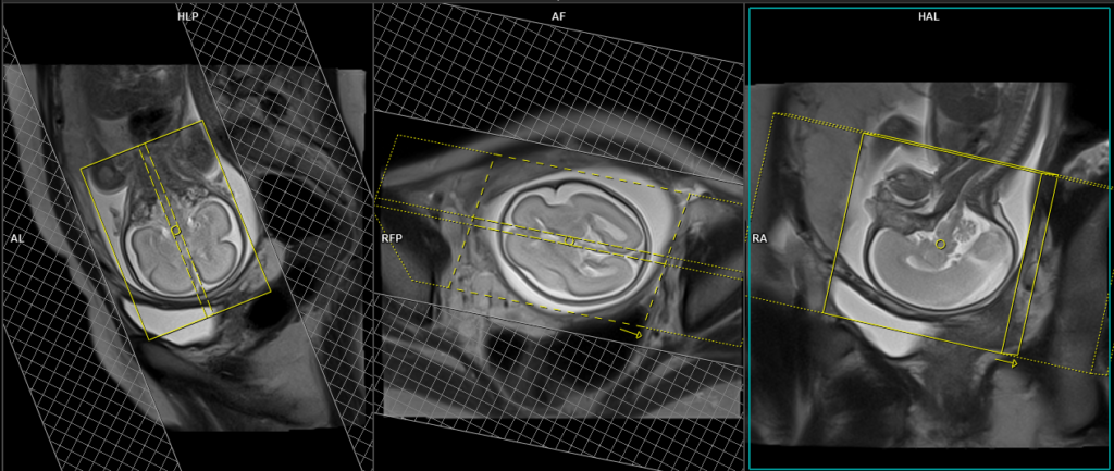
T2 HASTE sagittal localiser fetal brain
Plan the single-slice coronal localiser on the coronal uterus scans or sagittal localiser. Angle the position block perpendicular to the genu and splenium of the corpus callosum of the fetal head. Verify the positioning block in the other two planes to ensure proper alignment. In the axial scans, establish an appropriate angle running parallel to the midline of the fetal brain. The field of view (FOV) must be sufficiently large to cover the entire brain, typically ranging from 180 mm to 250 mm. It is important to include an appropriate amount of oversampling to prevent any wrap-around artifacts. These scans usually take approximately 1 to 2 seconds, and they can be performed either as a breath-hold or using the respiratory sensor.
Please note that the position of the fetal head may vary among different patients and during different trimesters. Therefore, it is crucial to plan the localiser according to the position of the fetal brain. The reference points mentioned in the planning process remain the same.

Parameters
TR 4-5 | TE 2-3 | FLIP 60 | NEX 1 | SLICE 5 MM | MATRIX 320×320 | FOV 400-480 | PHASE A>P | OVERSAMPLE 10% | IPAT ON |
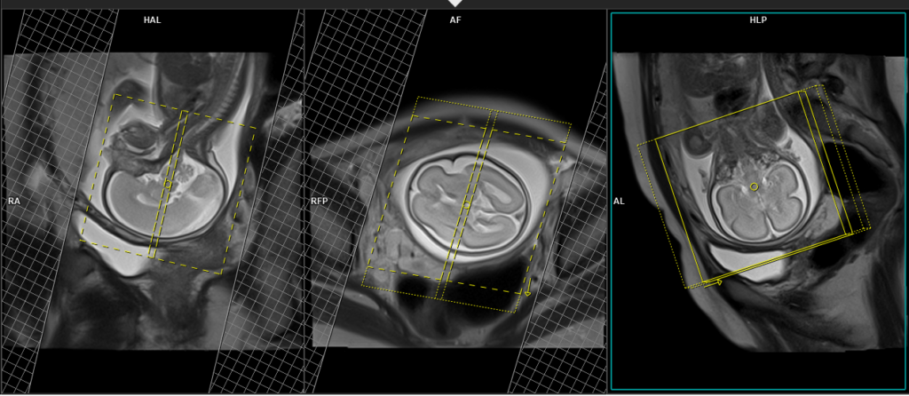
T2 HASTE axial 3mm fetal brain
Plan the axial scans on the sagittal localiser . Angle the position block parallel to the genu and splenium of the corpus callosum of the fetal head. Verify the positioning block in the other two planes to ensure proper alignment. In the coronal localizer, establish an appropriate angle running perpendicular to the midline of the fetal brain. The number of slices should be sufficient to cover the entire brain, ranging from the vertex to the line of the foramen magnum. The field of view (FOV) must be large enough to cover the entire brain, typically ranging from 180 mm to 220 mm. However, it is important to note that these scans usually take approximately 30 to 35 seconds, which can be challenging for a pregnant woman to hold her breath. To address this issue, we perform the scan under respiratory gating.
Parameters
TR 4-5 | TE 2-3 | FLIP 60 | NEX 1 | SLICE 5 MM | MATRIX 320×320 | FOV 400-480 | PHASE A>P | OVERSAMPLE 10% | IPAT ON |
T1 tse/ T1 VIBE axial 3mm breath hold fetal brain
Plan the axial scans on the sagittal localiser . Angle the position block parallel to the genu and splenium of the corpus callosum of the fetal head. Verify the positioning block in the other two planes to ensure proper alignment. In the coronal localiser, establish an appropriate angle running perpendicular to the midline of the fetal brain. The number of slices should be sufficient to cover the entire brain, ranging from the vertex to the line of the foramen magnum. The field of view (FOV) must be large enough to cover the entire brain, typically ranging from 180 mm to 220 mm. However, it is crucial to understand that these scans typically last for approximately 15 to 20 seconds and cannot be performed under respiratory gating. Therefore, kindly request the patient to hold their breath during this short duration. Based on our experience, most patients are willing to comply with the breath-holding instructions for a scan of this duration.
Parameters
TR 4-5 | TE 2-3 | FLIP 60 | NEX 1 | SLICE 5 MM | MATRIX 320×320 | FOV 400-480 | PHASE A>P | OVERSAMPLE 10% | IPAT ON |
EPI DWI axial 3mm free berthing fetal brain
Plan the axial scans on the sagittal localiser . Angle the position block parallel to the genu and splenium of the corpus callosum of the fetal head. Verify the positioning block in the other two planes to ensure proper alignment. In the coronal localiser, establish an appropriate angle running perpendicular to the midline of the fetal brain. The number of slices should be sufficient to cover the entire brain, ranging from the vertex to the line of the foramen magnum. The field of view (FOV) must be large enough to cover the entire brain, typically ranging from 180 mm to 220 mm. EPI DWI free-breathing scans typically have a duration of around 60 to 90 seconds and are conducted while the patient breathes normally.
Parameters
TR 4-5 | TE 2-3 | FLIP 60 | NEX 1 | SLICE 5 MM | MATRIX 320×320 | FOV 400-480 | PHASE A>P | OVERSAMPLE 10% | IPAT ON |
T2 HASTE sagittal 3mm fetal brain
Plan the sagittal scans on the axial localiser, ensuring that the position block is angled parallel to the midline of the fetal brain. Verify the positioning block in the other two planes to ensure proper alignment. In the coronal localiser, establish an appropriate angle running parallel to the midline of the fetal brain. The number of slices should be sufficient to cover the entire brain, ranging from one temporal lobe to the other. The field of view (FOV) must be large enough to encompass the entire brain, typically ranging from 180 mm to 220 mm. However, it is important to note that these scans usually take approximately 30 to 35 seconds, which can be challenging for a pregnant woman to hold her breath. To address this issue, we perform the scan under respiratory gating.
Parameters
TR 4-5 | TE 2-3 | FLIP 60 | NEX 1 | SLICE 5 MM | MATRIX 320×320 | FOV 400-480 | PHASE A>P | OVERSAMPLE 10% | IPAT ON |
T2 HASTE coronal 3mm fetal brain
Plan the coronal scans on the sagittal localiser . Angle the position block perpendicular to the genu and splenium of the corpus callosum of the fetal head. Verify the positioning block in the other two planes to ensure proper alignment. In the axial localiser, establish an appropriate angle running perpendicular to the midline of the fetal brain. The number of slices slices should be sufficient to cover the entire brain from the frontal sinus to the line of the occipital protuberance. The field of view (FOV) must be large enough to cover the entire brain, typically ranging from 180 mm to 220 mm. However, it is important to note that these scans usually take approximately 30 to 35 seconds, which can be challenging for a pregnant woman to hold her breath. To address this issue, we perform the scan under respiratory gating.
Parameters
TR 4-5 | TE 2-3 | FLIP 60 | NEX 1 | SLICE 5 MM | MATRIX 320×320 | FOV 400-480 | PHASE A>P | OVERSAMPLE 10% | IPAT ON |