MRI VIBE/LAVA-XV/THRIVE/TIGRE
Volumetric Interpolated Breath-hold Examination (VIBE) is a radio-frequency-spoiled 3D gradient echo (GRE) sequence utilized in magnetic resonance imaging (MRI) to create T1-weighted three-dimensional images employing interpolation and/or partial Fourier techniques. A derivative of the FLASH sequence, VIBE allows dynamic and high-resolution imaging within a 30-second breath-hold. In comparison to FLASH, VIBE maintains similar image quality (signal-to-noise ratio, contrast-to-noise ratio) while enhancing slice-selective spatial resolution (2 to 3 mm). Particularly advantageous for simultaneously evaluating soft tissue and vasculature, VIBE stands as an advanced imaging tool.
The defining attribute of VIBE MRI lies in its capacity to amass volumetric data during a solitary breath-holding instance. This is vital in mitigating motion-related blurring, especially when imaging moving organs or during extended acquisition periods. VIBE tackles this challenge through specific methodologies:
Breath-holding: Patients are instructed to suspend breathing for a brief interval during the imaging process. This mitigates motion artifacts, ensuring the acquired images remain sharp and precise.
Interpolation: The “interpolated” element of VIBE involves filling gaps between captured slices to construct a comprehensive three-dimensional volume. This interpolation enhances the spatial resolution of the final image, yielding a more intricate and comprehensive depiction of the imaged region.
Volumetric Imaging: VIBE MRI acquires volumetric data in a single scan, presenting a three-dimensional rendering of the imaged area. This capability is especially advantageous for visualizing complex structures like organs with irregular surfaces or structures spanning multiple planes.
MRI VIBE Image Appearance
2Fluid-Containing Structures:
- Cerebrospinal Fluid (CSF): CSF, found in the ventricles of the brain and around the spinal cord, appears dark or black on VIBE MRI due to its long T1 relaxation time.
- Synovial Fluid: Fluid within joint spaces, known as synovial fluid, also appears dark due to its long T1 relaxation time.
Soft Tissues:
- Muscles: Muscles exhibit intermediate signal intensity, appearing gray on VIBE images. The signal intensity can vary based on factors such as fat content and muscle type.
- Fat: Fat-rich tissues appear bright or white on VIBE MRI due to their short T1 relaxation time. This is particularly evident in subcutaneous fat and bone marrow.
Organs:
- Liver: The liver typically exhibits moderate signal intensity, appearing intermediate between dark and bright due to its mixed composition of blood vessels, parenchyma, and fat content.
- Spleen: The spleen often appears darker than the liver due to its higher iron content and relatively low fat content.
- Kidneys: Renal parenchyma usually appears gray, while renal pelvis and collecting system may appear darker due to fluid content.
Blood Vessels:
- Blood in Arteries and Veins: Blood flowing within arteries and veins appears as a flowing dark signal within bright vessel structures on VIBE MRI, thanks to the flow-related enhancement techniques used. Ocationaly the slow moving blood can apper bright on VIBE images
Bone and Bone Marrow:
- Bone: Bones appear dark or black on VIBE MRI due to their short T2 relaxation time and minimal water content.
- Bone Marrow: Depending on the composition (red vs. yellow marrow), bone marrow may appear with varying signal intensities.
pathology appearance on VIBE images
Hemorrhage:
- Acute Hemorrhage: Acute bleeding can appear as regions of high signal intensity on VIBE MRI, often due to the presence of paramagnetic substances like deoxyhemoglobin.
- Subacute Hemorrhage: Subacute hemorrhage may show varying signal intensities, with a mix of bright and intermediate signals as the blood components change over time.
Tumors:
- Solid Tumors: Solid tumors can appear as regions with altered signal intensities compared to surrounding tissues. This alteration may be due to differences in cellularity, vascularity, and water content.
- Contrast Enhancement: VIBE MRI can highlight enhanced regions within tumors, indicating areas of increased blood supply, potentially aiding in tumor characterization.
Cysts:
- Simple Cysts: Fluid-filled simple cysts may appear as regions of low signal intensity on VIBE MRI due to the lack of cellular content. The cystic fluid may exhibit low signal similar to other fluid-containing structures.
Inflammation:
- Inflammatory Processes: Areas of inflammation or edema may show low signal intensity on VIBE MRI due to increased water content in affected tissues.
Ischemic Changes:
- Infarction: Ischemic regions, such as those following a stroke, may exhibit altered signal intensity on VIBE MRI depending on the extent of tissue damage and edema.
Vascular Anomalies:
- Arteriovenous Malformations (AVMs): AVMs can be visualized as areas with abnormal blood vessel connections and altered signal intensity on VIBE MRI, often exhibiting enhanced vascularity.
Fatty Infiltration:
- Liver Steatosis: Fatty liver can appear brighter than normal liver tissue due to the increased fat content, resulting in higher signal intensity on VIBE MRI.
Calcifications:
- Calcified Lesions: Areas of calcification, such as in certain tumors or atherosclerotic plaques, may appear dark on VIBE MRI due to their minimal water content.
Lymphadenopathy:
- Enlarged Lymph Nodes: Enlarged lymph nodes may exhibit altered signal intensity on VIBE MRI, indicating potential changes in tissue composition and vascularity.
Degenerative Changes:
- Osteoarthritis: Degenerated joints and cartilage can exhibit signal changes on VIBE MRI due to altered tissue properties and potential joint effusion.
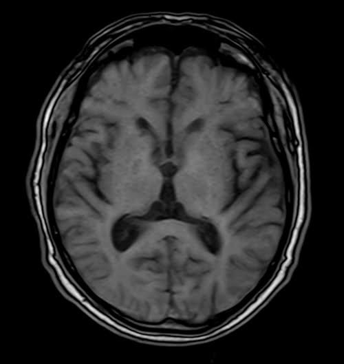
Tissues and their VIBE appearance
Bone marrow : – equal to or higher than that of muscle (fatty marrow is bright)
Spleen : bright gray( darker than liver)
Muscles :- gray
Moving blood : – gray
White matter : – whiter
Liver : bright gray
Gray matter : – gray
Fluids : – dark
Bone : – dark
Fat : – bright
Air : – dark
Use
- Useful for abdominal imaging
- Useful for small bowel and MR colonoscopy imaging
- Useful for chest imaging
- Useful for brain imaging (to acquire fast scans in uncooperative patients)
- Very useful for adrenal imaging
- Useful for liver imaging
Fat-saturated VIBE sequences are commonly employed in abdominal imaging, except for cases involving the adrenal gland, lipoma, or fatty liver imaging.
VIBE axial sequence used in uncooperative patients brain imaging
VIBE axial sequence used in adrenal gland imaging

VIBE coronal sequence used in adrenal gland imaging

VIBE axial sequence used in prostate imaging
