


Joseph Fourier

The origins of NMR (now known as MRI) trace back to the work of a French mathematician named Jean Baptiste Joseph Fourier (1768-1830). Fourier’s contributions to the field involved the development of a mathematical technique to study the transfer of heat between solid objects. This breakthrough eventually led to the advancement of phase and frequency signal processing in NMR, enabling rapid analysis in the field.

The unit of measurement for magnetic field strength, known as the Tesla, is named after Nikola Tesla, a Serbian inventor (1856-1943) who made significant contributions to the field of electromagnetism. One of his notable discoveries was the rotating magnetic field. Tesla’s work revolutionized the understanding and application of magnetic fields, laying the foundation for numerous technological advancements in the field of electricity and magnetism. His contributions continue to impact various industries, including the development of Magnetic Resonance Imaging (MRI) technology.
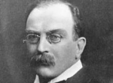
Sir Joseph Larmor (1857–1942), an accomplished Irish physicist, made significant contributions to the field of physics. Among his notable achievements, Larmor devised a method for calculating the rate at which energy is emitted by an accelerated electron. Additionally, he provided an explanation for the phenomenon of spectrum line splitting when subjected to a magnetic field. Larmor’s profound impact extends to the realm of Nuclear Magnetic Resonance (NMR), where he is renowned for formulating the “Larmor equation.” This equation states that the frequency of precession of the nuclear magnetic moment (ω) is directly proportional to the product of the magnetic field strength (B0) and the gyromagnetic ratio (γ), expressed as ω = γB0. Larmor’s pioneering work continues to underpin fundamental principles in the study of magnetic resonance.


In 1971, Raymond Damadian, a researcher from Downstate Medical Center in New York, conducted a groundbreaking study on the measurement of T1 and T2 relaxation times in rat tissues. His research focused on comparing normal tissue with cancerous tissue, and he made a significant discovery. Damadian found that normal tissue exhibited shorter relaxation times compared to tumor tissue. This observation marked an important milestone in the field of medical imaging and laid the foundation for the development of Magnetic Resonance Imaging (MRI) as a diagnostic tool. Damadian’s pioneering work provided crucial insights into the differences between healthy and diseased tissues, leading to the advancement of medical imaging techniques and their application in the detection and characterization of various conditions, including cancer. His contributions to the field of MRI have had a profound impact on modern healthcare, revolutionizing the way we diagnose and understand diseases.

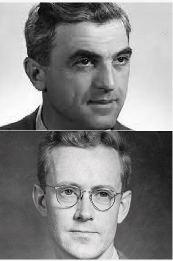
In 1974, Paul C. Lauterbur, a professor of chemistry and radiology at New York University, and Peter Mansfield from the Department of Physics at the University of Nottingham in England, made groundbreaking advancements in the field of magnetic resonance. Independently of each other, they described the utilization of magnetic field gradients to spatially localize NMR signals. Their remarkable discoveries formed the foundation for the revolutionary technology known as Magnetic Resonance Imaging (MRI).
As a result of their extraordinary contributions, Paul C. Lauterbur and Peter Mansfield were jointly awarded the Nobel Prize in Physiology or Medicine in 2003. This prestigious recognition served to honor their exceptional achievements and their profound impact on the field of medical imaging. Their work revolutionized diagnostic medicine and opened up new possibilities for non-invasive visualization of the human body.n 1974, Paul C. Lauterbur, a professor of chemistry and radiology at New York University and Peter Mansfield from the department of physics at the Nottingham University England independent of each other, described the use of magnetic field gradients for spatial localization of NMR signals. These discoveries led to the foundation for Magnetic Resonance Imaging (MRI). For this discovery, Lauterbur and Mansfield were awarded with the nobel prize for physiology or medicine in 2003.
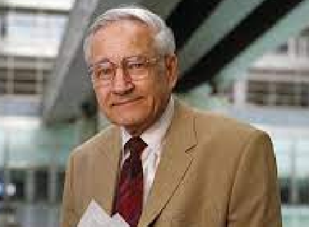
In 1975, a Swiss physical chemist Richard Ernst described the use of Fourier transform of phase and frequency encoding to reconstruct 2D images. For this discovery he was awarded with the nobel prize for chemistry in 1991.In 1975, Richard Ernst, a Swiss physical chemist, made a groundbreaking contribution to the field by introducing the concept of using Fourier transform of phase and frequency encoding for the reconstruction of two-dimensional (2D) images. This innovative technique revolutionized the field of medical imaging, particularly Magnetic Resonance Imaging (MRI). Ernst’s pioneering work opened up new possibilities for high-resolution imaging and significantly advanced the capabilities of MRI technology.
Due to the immense impact of his discovery, Richard Ernst was honored with the Nobel Prize in Chemistry in 1991. This prestigious recognition not only highlighted the significance of his contribution to the scientific community but also underscored the vital role of MRI in medical diagnostics and research. Richard Ernst’s innovative techniques continue to shape the field of MRI and have paved the way for further advancements in imaging technology.

Later in 1975, Peter Mansfield and Andrew Maudsley proposed a line scan technique, which led to the first cross sectional imaging of human anatomy (cross section through a finger). In 1978, Hugh Clow and Ian R. Young worked at a British company called EMI, created the first transverse NMR image through a human head.In 1975, Peter Mansfield and Andrew Maudsley made significant contributions to the field of medical imaging. They introduced a line scan technique that revolutionized the way we visualize human anatomy. This technique enabled the first-ever cross-sectional imaging of the human body, starting with a cross-section through a finger. Their pioneering work laid the foundation for the development of advanced imaging modalities like Magnetic Resonance Imaging (MRI).
Three years later, in 1978, Hugh Clow and Ian R. Young, who were working at a British company called EMI, achieved another groundbreaking milestone in medical imaging. They successfully created the first transverse Nuclear Magnetic Resonance (NMR) image of a human head. This achievement opened up new possibilities for non-invasive imaging of the brain and other internal structures, providing valuable insights for medical diagnosis and research.
The contributions of Peter Mansfield, Andrew Maudsley, Hugh Clow, and Ian R. Young have had a profound impact on the field of medical imaging. Their innovative techniques and advancements have paved the way for the widespread use of MRI, revolutionizing diagnostic medicine and greatly enhancing our understanding of the human body.