Thymus (Mediastinum) MRI
Indications for chest MRI scan
- Thymic hyperplasia
- Thymic cysts
- Lymphoma
- Mediastinal mass
- benign and malignant neoplasms
Contraindications
- Any electrically, magnetically or mechanically activated implant (e.g. cardiac pacemaker, insulin pump biostimulator, neurostimulator, cochlear implant, and hearing aids)
- Intracranial aneurysm clips (unless made of titanium)
- Pregnancy (risk vs benefit ratio to be assessed)
- Ferromagnetic surgical clips or staples
- Metallic foreign body in the eye
- Metal shrapnel or bullet
Patient preparation for Thymus MRI scan
- A satisfactory written consent form must be taken from the patient before entering the scanner room
- Ask the patient to remove all metal objects including keys, coins, wallet, cards with magnetic strips, jewellery, hearing aid and hairpins
- Ask the patient to undress and change into a hospital gown
- Instruct the patient to hold their breath for the breath hold scans and breathe gently for the gated scans (it is advisable to coach the patient two to three times before starting the scan)
- An intravenous line must be placed with extension tubing extending out of the magnetic bore
- If possible provide a chaperone for claustrophobic patients (e.g. relative or staff )
- Offer earplugs or headphones, possibly with music for extra comfort
- Explain the procedure to the patient
- Instruct the patient to keep still
- Note down the weight of the patient
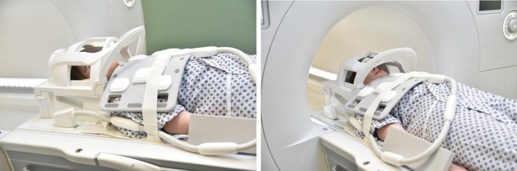
Positioning for Thymus MRI scan
- Position the patient in supine position with head pointing towards the magnet (head first supine)
- Position the patient over the spine, head and neck coil and place the neck and body\large flex coil over the neck and upper chest (nose tip down to xiphoid process)
- Securely tighten the body coil using straps to prevent respiratory artefacts
- Give cushions under the head and legs for extra comfort
- Center the laser beam localizer over the mid-sternum.
Recommended Thymus MRI scan Protocols and Planning
localiser
To localize and plan the sequences, it is essential to acquire a three-plane T2 HASTE localizer initially. These fast single-shot localizers have an acquisition time of under 25 seconds and are highly effective in accurately localizing chest and abdominal structures.

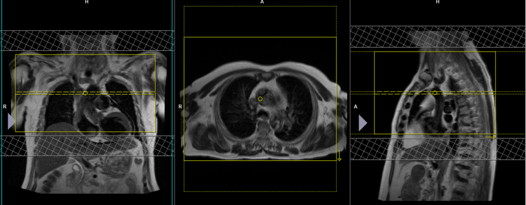
T2 tse\HASTE Multiple breath-hold axial 6mm
Plan the axial slices on the coronal image and position the block horizontally across the chest as shown. Verify the positioning in the other two planes. Establish an appropriate angle in the sagittal plane by aligning it vertically across the chest. The slices must cover the entire chest from the mid-neck down to the aortic valve. Instruct the patient to hold their breath during image acquisition. The parallel acquisition technique (IPAT/SENSE) can be used to reduce the scan time. Additionally, adding saturation bands on the top and bottom of the axial block will help reduce arterial pulsation and breathing artifacts.
Parameters
TR 5000-7000 | TE 90 | FLIP 150 | NXA 1 | SLICE 6 MM | MATRIX 256×256 | FOV 350 | PHASE A>P | OVERSAMPLE 50% | IPAT ON |
T1 DIXON 3D breath hold coronal 3mm SFOV
Plan the coronal slices on the axial scan and position the block horizontally across the chest as shown. Verify the positioning in the other two planes. Establish an appropriate angle in the sagittal plane by aligning it vertically across the chest. Ensure that the slices adequately cover the entire mediastinum from the anterior chest wall to the vertebral body. To prevent wrap-around artifacts, phase oversampling and slice oversampling must be used. Instruct the patient to hold their breath during image acquisition. In our department, we instruct patients to take two deep breaths before giving the ‘breathe in and hold’ instruction.

Parameters
TR 6-7 | TE 2.39 4.77 | FLIP 10 | NXA 1 | SLICE 3 MM | MATRIX 224×224 | FOV 200 | PHASE R>L | OVERSAMPLE 50% | BH YES |
T2 tse\HASTE Multiple breath-hold coronal 3mm SFOV
Plan the coronal slices on the axial scan and position the block horizontally across the chest as shown. Verify the positioning in the other two planes. Establish an appropriate angle in the sagittal plane by aligning it vertically across the chest. Ensure that the slices adequately cover the entire mediastinum, from the anterior chest wall to the vertebral body. To prevent wrap-around artifacts, phase oversampling must be used. Instruct the patient to hold their breath during image acquisition. In our department, we instruct patients to breathe in and out twice before giving the ‘breathe in and hold’ instruction.

Parameters
TR 4000-5000 | TE 90 | FLIP 150 | NXA 1 | SLICE 3 MM | MATRIX 224×224 | FOV 220 | PHASE R>L | OVERSAMPLE 50% | BH YES |
T1 DIXON 3D axial breath hold 3mm SFOV
Plan the axial slices on the coronal image and position the block horizontally across the chest as shown. Verify the positioning in the other two planes. Establish an appropriate angle in the sagittal plane by aligning it horizontally across the chest. Ensure that the slices adequately cover the entire mediastinum from the mid-neck to the aortic valve. To prevent wrap-around artifacts, phase oversampling and slice oversampling must be used. Instruct the patient to hold their breath during image acquisition. In our department, we instruct patients to breathe in and out twice before giving the ‘breathe in and hold’ instruction.

Parameters
TR 7-8 | TE 2.39 4.77 | FLIP 10 | NXA 1 | SLICE 3 MM | MATRIX 224×224 | FOV 200 | PHASE R>L | OVERSAMPLE 50% | BH YES |
T2 tse \HASTE Multiple breath-hold axial 3mm SFOV
Plan the axial slices on the coronal image and position the block horizontally across the chest as shown. Verify the positioning in the other two planes. Establish an appropriate angle in the sagittal plane by aligning it horizontally across the chest. Ensure that the slices adequately cover the entire mediastinum from the mid-neck to the aortic valve. To prevent wrap-around artifacts, phase oversampling must be used. Instruct the patient to hold their breath during image acquisition. In our department, we instruct patients to breathe in and out twice before giving the ‘breathe in and hold’ instruction.

Parameters
TR 4000-5000 | TE 90 | FLIP 150 | NXA 1 | SLICE 3 MM | MATRIX 224×224 | FOV 220 | PHASE A>P | OVERSAMPLE 30% | BH YES |
T2 tse fat sat\HASTE fat sat Multiple breath-hold axial 3mm SFOV
Plan the axial slices on the coronal image and position the block horizontally across the chest as shown. Verify the positioning in the other two planes. Establish an appropriate angle in the sagittal plane by aligning it horizontally across the chest. Ensure that the slices adequately cover the entire mediastinum from the mid-neck to the aortic valve. To prevent wrap-around artifacts, phase oversampling must be used. Instruct the patient to hold their breath during image acquisition. In our department, we instruct patients to breathe in and out twice before giving the ‘breathe in and hold’ instruction.
Parameters
TR 6000-8000 | TE 90 | fatsat ON | NXA 1 | SLICE 3 MM | MATRIX 208×208 | FOV 220 | PHASE A>P | OVERSAMPLE 30% | IPAT ON |
DWI epi 3 scan trace axial 3mm free breathing
Plan the axial slices on the coronal image and position the block horizontally across the chest as shown. Verify the positioning in the other two planes. Establish an appropriate angle in the sagittal plane by aligning it horizontally across the chest. Ensure that the slices adequately cover the entire mediastinum from the mid-neck to the aortic valve. To prevent wrap-around artifacts, phase oversampling must be used. Consider adding saturation bands at the top and bottom of the block to minimize artifacts caused by fat signal, arterial pulsation, and breathing

Parameters
TR 6000-7000 | TE 90 | IPAT ON | NEX 3 5 8 | SLICE 3 MM | MATRIX 192X192 | FOV 200-250 | PHASE R>L | GAP 10% | B VALUE 0 |
T2 tse \HASTE Multiple breath-hold sagittal 3mm SFOV
Plan the sagittal slices on the coronal image, positioning the block vertically across the chest as shown. Verify the positioning in the other two planes. Establish an appropriate angle in the axial plane by aligning it horizontally across the chest. The slices must cover the entire mediastinum from right to left. Instruct the patient to hold their breath during image acquisition. The parallel acquisition technique (IPAT/SENSE) can be used to reduce the scan time. Adding saturation bands will help reduce arterial pulsation and breathing artifacts.

Parameters
TR 6000-7000 | TE 90 | fatsat ON | NXA 1 | SLICE 6 MM | MATRIX 256×256 | FOV 350 | PHASE A>P | OVERSAMPLE 50% | IPAT ON |
For the post-contrast scans, use T1 vibe DIXON axial, coronal and sagittal sequences after the administration of IV gadolinium DTPA injection (copy the planning outlined above). The document below provides access to the recommended dosage of gadolinium DTPA injection, as advised by the manufacturer.