Penile MRI
Indications for penile mri scan
- Metastatic tumours of the urethra or penis
- Malignant tumours of the urethra or penis
- Benign tumours of the urethra or penis
- Tuberculosis of the urethra or penis
- Blunt penile or urethral trauma
- Penetrating penile injury
- Condyloma acuminata
- Postirradiation Injuries
- Penile fracture
Contraindications for penile mri scan
- Any electrically, magnetically or mechanically activated implant (e.g. cardiac pacemaker, insulin pump biostimulator, neurostimulator, cochlear implant, and hearing aids)
- Intracranial aneurysm clips (unless made of titanium)
- Pregnancy (risk vs benefit ratio to be assessed)
- Ferromagnetic surgical clips or staples
- Metallic foreign body in the eye
- Metal shrapnel or bullet
Patient preparation for penile mri scan
- A satisfactory written consent form must be taken from the patient before entering the scanner room
- Ask the patient to remove all metal objects including keys, coins, wallet, cards with magnetic strips, jewellery, hearing aid and hairpins
- Ask the patient to undress and change into a hospital gown
- Contrast injection risk and benefits must be explained to the before the scan
- Gadolinium should only be given to the patient if GFR is > 30
- If possible provide a chaperone for claustrophobic patients (e.g. relative or staff )
- Offer earplugs or headphones, possibly with music for extra comfort
- Explain the procedure to the patient
- Instruct the patient to keep still
- Note the weight of the patient
Positioning for penile mri scan
- Position the patient in supine position with head pointing towards the magnet (head first supine).
- Position the patient over the spine coil and place the body coil over the pelvis( iliac crest down to three inches below testicle)
- Securely tighten the body coil using straps to prevent respiratory artefacts
- Give a pillow under the head for extra comfort (do not give cushions under the legs )
- Centre the laser beam localiser over pubic symphysis

Recommended Penile MRI Protocols, Parameters, and Planning
Penile mri localiser
A three-plane HASTE localizer must be taken initially to localize and plan the sequences. These are fast single-shot localizers with under 25s acquisition time, which are excellent for localizing pelvic structures. Take at least 3-4 slices in all planes to get the best results.

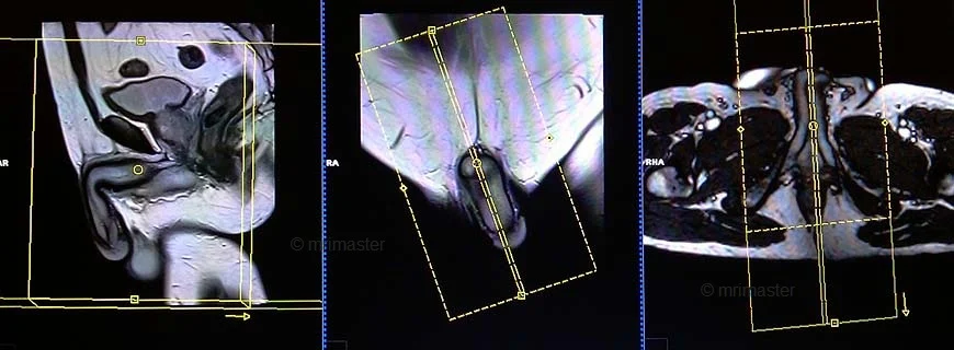
T2 tse sagittal 3mm SFOV
Plan the sagittal slices on the axial plane. Angle the positioning block parallel to the interpubic fibrocartilage and the anal canal (i.e. parallel to the penile base). Check the positioning block in the other two planes. An appropriate angle must be given in the coronal plane (parallel to the penis). Slices must be sufficient to cover the entire penis. The FOV must be big enough to cover the whole penis (normally 180mm-200mm).
Parameters
TR 3000-4000 | TE 100-120 | SLICE 3 MM | FLIP 130-150 | PHASE A>P | MATRIX 320X320 | FOV 270-300 | GAP 10% | NEX(AVRAGE) 3 |
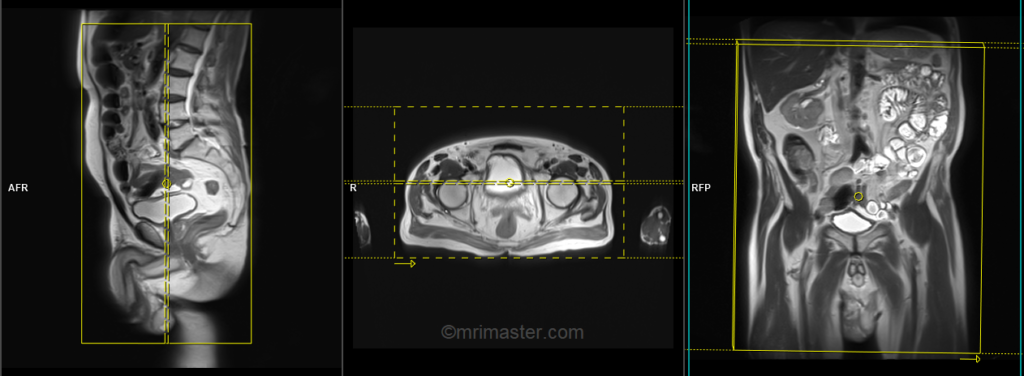
T2 tse coronal 5mm big FOV(For cancer imaging)
Plan the big FOV coronal slices on the axial plane and angle the positioning block parallel to the right and left hip joint. Check the positioning block in the other two planes. An appropriate angle must be given in the sagittal plane (vertically across the abdomen). The slices must be sufficient to cover the entire abdomen and pelvis from anterior to posterior. The FOV must be big enough to cover the abdomen and pelvis, typically ranging from 350mm to 400mm. Big FOV scans are only required for cancer imaging of the penis; there is no need to perform this sequence for penile fracture imaging.
Parameters
TR 4000-5000 | TE 100-120 | SLICE 5 MM | FLIP 130-150 | PHASE R>L | MATRIX 384X384 | FOV 350-400 | GAP 10% | NEX(AVRAGE) 2 |
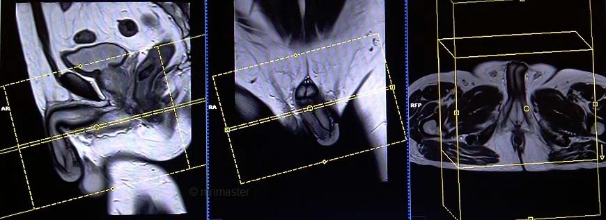
T2 stir axial 3mm SFOV
Plan the axial slices on the sagittal plane and angle the positioning block parallel to the penile base (i.e., perpendicular to the penile shaft and glans penis). Check the positioning block in the other two planes, ensuring an appropriate angle is given in the coronal plane (perpendicular to the penile shaft and glans penis). The slices must be sufficient to cover the entire penis from the prostate down to 1cm below the glans penis.
Parameters
TR 4000-5000 | TE 110 | FLIP 130 | NEX 4 | SLICE 3 MM | MATRIX 256X256 | FOV 180-200 | PHASE A>P | GAP 10% | TI 130 |
T1 tse axial 3mm SFOV
Plan the axial slices on the sagittal plane and angle the positioning block parallel to the penile base (i.e., perpendicular to the penile shaft and glans penis). Check the positioning block in the other two planes, ensuring an appropriate angle is given in the coronal plane (perpendicular to the penile shaft and glans penis). The slices must be sufficient to cover the entire penis from the prostate down to 1cm below the glans penis.
Parameters
TR 400-600 | TE 15-25 | SLICE 3 MM | FLIP 140 | PHASE A>P | MATRIX 2320X256 | FOV 180-200 | GAP 10% | NEX(AVRAGE) 3 |
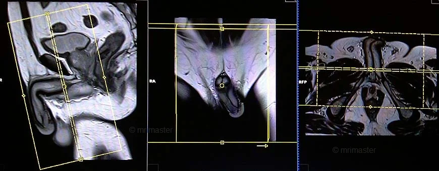
T2 stir coronal 3mm SFOV
Plan the coronal slices on the sagittal plane and angle the positioning block parallel to the penile shaft and glans penis. Check the positioning block in the other two planes. An appropriate angle must be given in the axial plane (perpendicular to the base of the penis). The slices must be sufficient to cover the entire penis from anterior to posterior.
Parameters
TR 4000-5000 | TE 110 | FLIP 130 | NEX 4 | SLICE 3 MM | MATRIX 256X256 | FOV 180-200 | PHASE R>L | GAP 10% | TI 150 |
T1 tse coronal 3mm SFOV
Plan the coronal slices on the sagittal plane and angle the positioning block parallel to the penile shaft and glans penis. Check the positioning block in the other two planes. An appropriate angle must be given in the axial plane (perpendicular to the base of the penis). The slices must be sufficient to cover the entire penis from anterior to posterior.
Parameters
TR 400-600 | TE 15-25 | SLICE 3 MM | FLIP 140 | PHASE R>L | MATRIX 256X256 | FOV 180-200 | GAP 10% | NEX(AVRAGE) 4 |