MRI Susceptibility weighted imaging (SWI)
Susceptibility Weighted Imaging (SWI) is an advanced MRI (Magnetic Resonance Imaging) technique that exploits the magnetic susceptibility differences between various tissue types and blood oxygen levels to provide detailed visualization of venous structures, microbleeds, and other vascular abnormalities. Compared to conventional MRI sequences, SWI offers an unparalleled ability to reveal subtle vascular and hemorrhagic phenomena that are often undetected in standard imaging techniques.
Physics of SWI
The underlying principle of SWI (Susceptibility Weighted Imaging) revolves around the magnetic susceptibility differences among various tissues. When tissues of differing magnetic susceptibilities are exposed to a magnetic field, they generate minor local magnetic field variations. These fluctuations result in phase differences in MRI signals. While traditional MRI sequences often discard this phase information, SWI leverages it to produce an image.
The process by which SWI forms an image is as follows:
Magnetic Field Inhomogeneities: The presence of substances like deoxygenated blood or iron in the brain distorts the local magnetic field. This distortion leads to both magnitude (signal intensity) and phase changes in the MRI signal.
Phase Images: SWI sequences capture both magnitude and phase data. This phase data is subsequently unwrapped and high-pass filtered to eliminate global phase variations, emphasizing local differences.
Combining Magnitude and Phase Data: The phase information is combined with the magnitude data to enhance contrast based on magnetic susceptibility differences. The result is an image with heightened contrast in regions showcasing susceptibility variations.
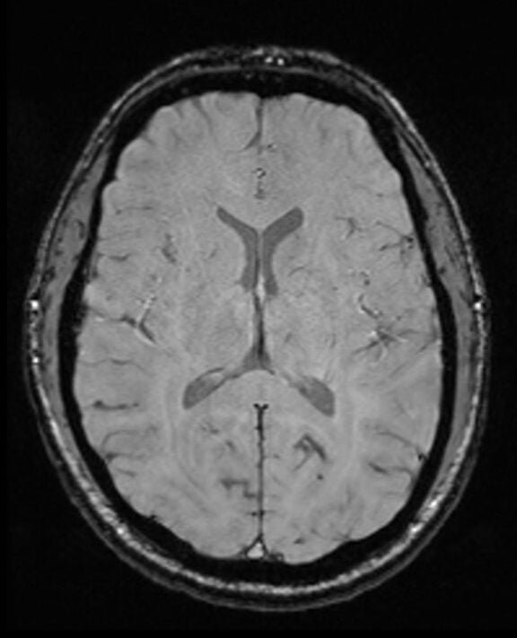
SWI: Magnitude image
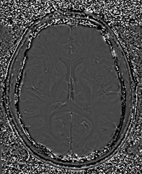
SWI: Phase images
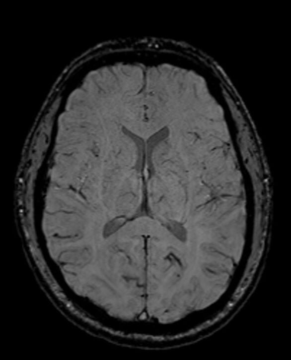
SWI: Magnitude + Phase image
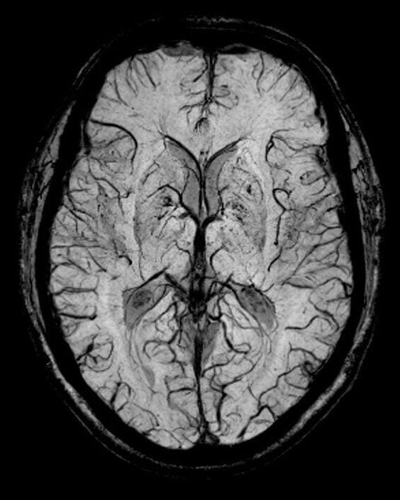
SWI: (Magnitude + Phase) MIP image
Image appearance of SWI MRI
In SWI images, veins appear distinctly black due to the deoxygenated blood they contain. Brain tissue generally displays an intermediate brightness, providing a clear contrast against the backdrop. Cerebrospinal fluid (CSF) ranges from intermediate to dark, enhancing visibility of surrounding structures. Both bone and calcifications appear dark, making them easy to differentiate from soft tissues. Fat and muscle tissues typically present with an intermediate signal.
Applications of SWI
Visualization of Venous Structures: SWI provides a clear depiction of venous vasculature, particularly small veins that might not be well-visualized using other MRI techniques.
Detection of Microbleeds: Brain microbleeds, which are small chronic brain hemorrhages, are better visualized with SWI due to the susceptibility effect of blood breakdown products.
Traumatic Brain Injury: SWI can help detect shearing injuries, microhemorrhages, and other vascular injuries in patients with traumatic brain injury.
Stroke: In cases of stroke, SWI can provide valuable information about the presence and extent of hemorrhage or the pattern of venous drainage.
Tumors: SWI can help in characterizing certain tumors based on the presence of hemorrhage, calcification, or vascular patterns.
Neurodegenerative Diseases: In conditions like Parkinson’s disease or multiple sclerosis, SWI can be used to visualize iron deposition in the brain, providing valuable diagnostic and research insights.
Arteriovenous Malformations (AVMs): SWI can highlight the abnormal blood vessels and associated hemorrhages in AVMs.
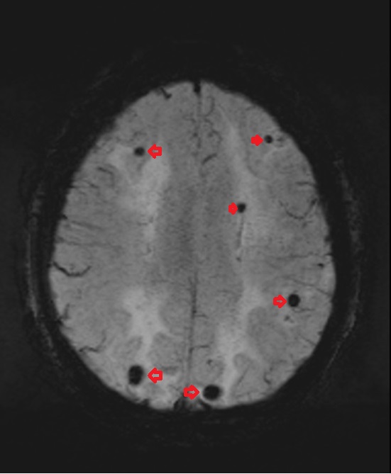
microbleed appearance in SWI

microbleed appearance in SWI
References
- Halefoglu, A. M., & Yousem, D. M. (2018). Susceptibility weighted imaging: Clinical applications and future directions. World Journal of Radiology, 10(4), 30–45.
Haacke EM, Xu Y, Cheng YC, et al. Susceptibility weighted imaging (SWI). Magn Reson Med. 2004;52(3):612-618.
Deistung A, Schweser F, Reichenbach JR. Overview of quantitative susceptibility mapping. NMR Biomed. 2017;30(4):e3569.
Mittal S, Wu Z, Neelavalli J, Haacke EM. Susceptibility-weighted imaging: technical aspects and clinical applications, part 2. AJNR Am J Neuroradiol. 2009;30(2):232-252.
Sehgal V, Delproposto Z, Haacke EM, et al. Clinical applications of neuroimaging with susceptibility-weighted imaging. J Magn Reson Imaging. 2005;22(4):439-450.