MRI Concatenations
Concatenations
Concatenations serve as a quantifiable parameter for assessment. They represent a technique for uniformly distributing slices across multiple TRs, and occasionally necessitate operator intervention for manipulation
Relevant Applications of Concatenations
Enhancement of Tissue Contrast: The quantity of slices required to encompass an anatomical area is constrained by the TR. In scenarios where the inclusion of additional slices would lengthen the TR, a strategic augmentation of concatenations permits a reduction in TR. This proves particularly beneficial during T1 scans.
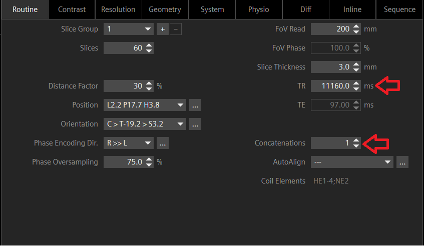
MRI Concatenations 1 with a high TR.

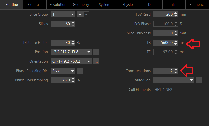
MRI Concatenations 2 with appropriate TR.

Optimization of Breath-hold Scans: Elevating the concatenation factor contributes to the curtailment of scan durations during breath-hold acquisitions. This approach leads to a greater frequency of shorter breath-holds, thereby enhancing patient tolerance.
Mitigation of Signal Cross-talk: Leveraging concatenations proves advantageous in forestalling signal cross-talk within images characterized by minimal or non-essential slice gaps, intervals, or distances. Opting for an interleaved slice sequence, wherein alternate slices are acquired (1, 3, 5 and 2, 4, 6), effectively averts cross-excitation.
Key Considerations:
Impact on Scan Duration: The duration of scans exhibits a direct correlation with concatenation levels. Doubling the concatenation factor results in a commensurate doubling of scan time. Consequently, this approach might be less efficient for expanding anatomical coverage, prompting a preference for augmenting TR as an alternative strategy.
Influence of Motion: In cases where patients are unable to maintain immobility, the acquired slice sets might not faithfully represent the patient’s positioning. Consequently, the potential exists for the oversight of pathologies. This phenomenon is often observable during the review of slices on PACS, where apparent “jumping” between slices occurs.
References
- Haacke, E. M., Brown, R. W., Thompson, M. R., & Venkatesan, R. (2015). Magnetic resonance imaging: Physical principles and sequence design. John Wiley & Sons.
- Bernstein, M. A., King, K. F., & Zhou, X. J. (2004). Handbook of MRI pulse sequences. Academic Press.
- Cline, H. E. (1990). Nuclear magnetic resonance imaging: physical principles and sequence design. Radiology, 177(1), 239-247.


