QISS (Non-Contrast) MRA Lower Limb
QISS MRI technology Introduction
QISS MRA (Quiescent-Inflow Single-Shot Magnetic Resonance Angiography) is a non-enhanced technique used in magnetic resonance angiography to evaluate peripheral arterial disease (PAD) in patients with impaired renal function. This technique is particularly useful because it avoids the risk of nephrogenic systemic fibrosis associated with contrast agents. QISS MRA is one of the newer non-enhanced techniques, along with fresh blood imaging and flow-sensitive dephasing.
The QISS MRA technique employs an undersampled Cartesian k-space trajectory combined with parallel imaging to reduce echo train length. However, traditional Cartesian undersampling factors larger than two to four usually result in poor image quality at 1.5 Tesla. To overcome this limitation, QISS MRA utilizes a radial k-space trajectory, which allows for high undersampling factors without sacrificing spatial resolution. The data acquired through QISS MRA is sparse, minimizing radial streak artifacts.
In QISS MRA, sparse images of arteries are generated by combining in-plane saturation and chemical shift-dependent radiofrequency fat saturation techniques. This approach enables high undersampling factors to accelerate data acquisition. The method has been optimized for multi-slice QISS MRA, allowing for the acquisition of data from multiple slices within each RR interval, reducing scan duration.
The implementation of QISS MRA involves the application of in-plane saturation and venous inferior tracking saturation pulses at a user-selected time delay after the R-wave. The readout is acquired during the quiescent interval, which ensures data acquisition during slow diastolic flow. The technique has been tested on healthy subjects and patients with PAD.
Compared to contrast-enhanced MRA, QISS MRA provides an accurate non-contrast alternative for visualizing arterial disease, especially in patients where contrast agents are contraindicated or those with renal dysfunction. QISS MRA is considered an easy-to-handle approach for imaging peripheral arteries with minimal requirements for further adjustments. It offers the potential to improve patient safety by reducing the need for contrast agents and associated risks.
Indications for lower limb qiss mra
- Peripheral arterial occlusive disease
- Post operative and stent evaluation
- Arteriovenous malformation
- Chronic critical ischemia
- Chronic arterial disease
- Acute arterial disease
- Aortoiliac disease
- Embolic disease
Contraindications
- Any electrically, magnetically or mechanically activated implant (e.g. cardiac pacemaker, insulin pump biostimulator, neurostimulator, cochlear implant, and hearing aids)
- Intracranial aneurysm clips (unless made of titanium)
- Pregnancy (risk vs benefit ratio to be assessed)
- Ferromagnetic surgical clips or staples
- Metallic foreign body in the eye
- Metal shrapnel or bullet
Patient preparation for qiss mra
- A satisfactory written consent form must be taken from the patient before entering the scanner room
- Ask the patient to remove all metal objects including keys, coins, wallet, cards with magnetic strips, jewellery, hearing aid and hairpins
- Ask the patient to undress and change into a hospital gown
- Ensure proper connection of ECG electrodes and position the ECG module towards the head.
- If possible provide a chaperone for claustrophobic patients (e.g. relative or staff )
- Offer earplugs or headphones, possibly with music for extra comfort
- Explain the procedure to the patient
- Instruct the patient to keep still
- Note the weight of the patient
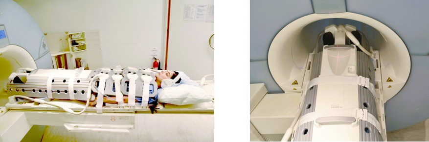
Positioning for qiss mra
- Position the patient over the spine coil and place the peripheral coil over the legs, and the two body coils over the abdomen and chest.
- Connect the ECG electrodes as specified above and in accordance with the specific manufacturer’s instructions.
- Position the ECG module towards the head.
- Securely tighten the body coil using straps to prevent respiratory artefacts
- Provide cushions under the head for extra comfort
- Centre the laser beam localiser over mid lower leg
Recommended QISS MRA Protocols, Parameters, and Planning
QISS MRA localiser
To ensure precise localization and sequence planning, it is recommended to start with a 3D localizer scan. These rapid, low-resolution volumetric localizers have an acquisition time of less than 25 seconds, making them ideal for accurately identifying lower limb structures.

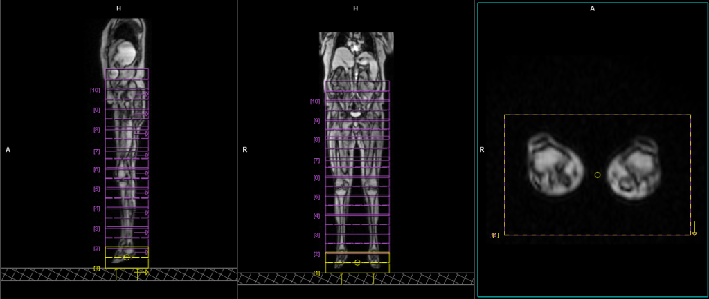
QISS TRUFISP AXIAL ECG gated 3MM
Plan the axial slices on the sagittal localizer and orient the position block horizontally across the pelvis and lower limb. Verify the positioning block in the other two planes. Ensure an appropriate angle is set in the coronal plane, horizontally spanning the pelvis and lower limb. The number of slices should sufficiently cover the arterial supply of the lower limb, from the renal arteries down to the plantar aspect of the foot. he QISS TRUFISP is performed as multy slice axial scans with 20% overlap between the blocks. Usually, 10 to 12 blocks are required to cover the pelvis and lower limb. Please add additional blocks for tall patients. Use the angiography composing function in the scanner to stich the images so it can be MIP as a single block. Use a saturation band at the bottom of the block to suppress the veinous signal.
Parameters
TR 811 | TE 2.02 | TI 345 | NEX 1 | SLICE 3 MM | MATRIX 400×400 | FOV 400 | PHASE A>P | ECG GATE YES | IPAT ON |
Post processing of QISS MRA data
MPR (Multi-Planar Reconstruction)
MPR (Multi-Planar Reconstruction) reconstruction is a technique used in MRI (Magnetic Resonance Imaging) to generate images in multiple planes from a set of acquired image data. It allows visualization of anatomical structures from different perspectives, such as axial, sagittal, and coronal planes, which are orthogonal to each other. MPR reconstruction involves extracting image data from the original three-dimensional volume dataset and reformats it into two-dimensional images along specific planes.

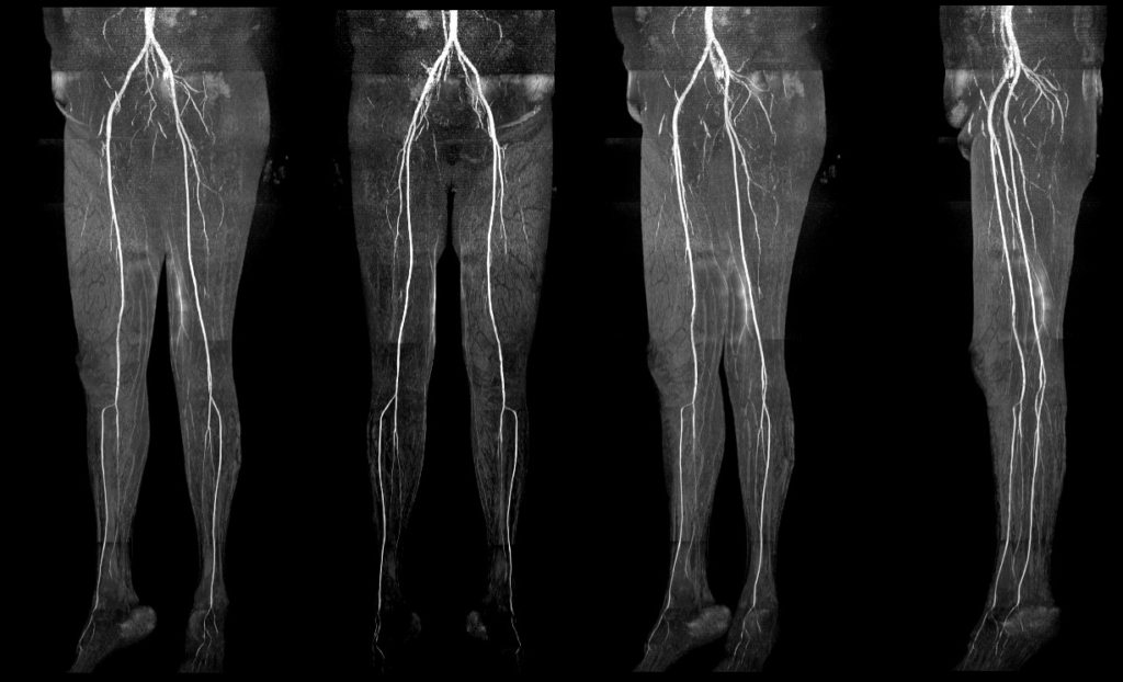
MIP (Maximum Intensity Projection)
MIP (Maximum Intensity Projection) reconstruction is a technique used in MRI (Magnetic Resonance Imaging) to create a two-dimensional image that displays the maximum intensity along a chosen projection path. It involves projecting the highest signal intensity voxel from each slice along a specific viewing direction onto a single image plane. MIP reconstructions are commonly used in vascular imaging to enhance the visualization of blood vessels and highlight areas of high signal intensity. This technique allows for a comprehensive overview of the vasculature and aids in identifying abnormalities or areas of interest in a three-dimensional volume dataset.