Bounce Point Artifact
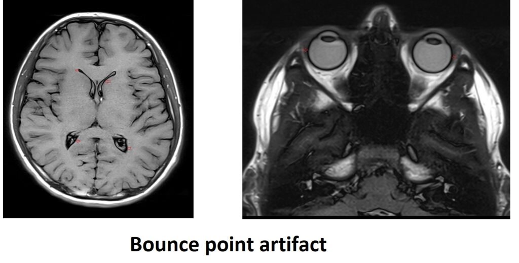
The bounce point artifact occurs in inversion recovery sequences along tissue boundaries. In an IR sequence an initial RF pulse flips the magnetisation through 180° and tissues relax back at their specific T1 rates passing through null point where there is no signal. If a TI is chosen which is intermediate between the null (or ‘bounce’) point of two different tissues, then the signal intensity of each tissue at TI will be equal but opposite (positive and negative) and the signals will cancel each other out.
The effect of the bounce point artifact on the image is the appearance of a black line at the border between two tissues, such as fat and muscle, or brain and cerebrospinal fluid (CSF). This artifact is also known as the ‘india ink’ artifact due to its resemblance to lines drawn with black ink.
Here are some strategies to minimize or avoid bounce point artifact :
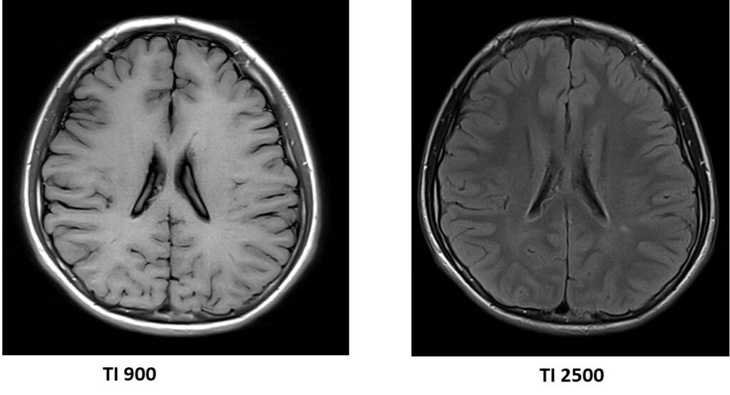
Adjust the inversion time (TI): Since the bounce point artifact occurs when the TI is chosen to be intermediate between the null points of two tissues, selecting a different TI can help mitigate the artifact. By carefully selecting the TI, one can avoid the cancellation of signals and achieve better tissue contrast without the artifact.
Use alternative imaging sequences: In some cases, using alternative imaging sequences, such as spin echo or fast spin echo, can help minimize the bounce point artifact.
References:
Saranathan, M., Worters, P. W., Rettmann, D. W., Winegar, B., & Becker, J. (2017). Physics for Clinicians: Fluid-Attenuated Inversion Recovery (FLAIR) and Double Inversion Recovery (DIR) Imaging. J. MAGN. RESON. IMAGING, 46, 1590–1600.
Saranathan, M., Worters, P.W., Rettmann, D.W., Winegar, B., & Becker, J. (Year). IPhysics for Clinicians: Fluid-Attenuated Inversion Recovery (FLAIR) and Double Inversion Recovery (DIR) Imaging.