Trigeminal Nerve(CN V) MRI
Indications for Trigeminal nerve MRI scan
- Trigeminal neuralgia
- Nerve schwannoma
- Hemifacial spasm
- Tic douloreux
Trigeminal nerve anatomy
The trigeminal nerve is one of the 12 cranial nerves and is responsible for transmitting sensory information from the face to the brain. It is the largest cranial nerve and has both sensory and motor functions. The trigeminal nerve is divided into three main branches: the ophthalmic nerve (V1), the maxillary nerve (V2), and the mandibular nerve (V3). Let’s explore each branch in more detail:
Ophthalmic Nerve (V1):
The ophthalmic nerve is the smallest branch and provides sensory innervation to the upper part of the face, including the forehead, scalp, upper eyelid, and the front part of the scalp.
It also carries sensory information from the cornea, conjunctiva, and the nasal cavity.
Maxillary Nerve (V2):
The maxillary nerve supplies sensory innervation to the middle part of the face, including the lower eyelid, upper lip, cheek, and part of the nasal cavity.
It also provides sensory input from the maxillary teeth, palate, sinuses, and the meninges of the middle cranial fossa.
Mandibular Nerve (V3):
The mandibular nerve is the largest branch of the trigeminal nerve and has both sensory and motor functions.
Sensory innervation includes the lower lip, chin, lower teeth, part of the cheek, and the anterior two-thirds of the tongue.
Motor fibers of the mandibular nerve control the muscles involved in chewing (mastication).
Trigeminal neuralgia
Trigeminal neuralgia is a debilitating condition characterized by severe facial pain originating from the trigeminal nerve. The intense, electric shock-like pain episodes can significantly impact a person’s daily life, causing difficulty with eating, speaking, and even simple facial movements. MRI plays a crucial role in the diagnosis of trigeminal neuralgia as it helps identify the underlying causes. It can detect potential compressions of the trigeminal nerve by nearby blood vessels, tumors, or structural abnormalities. MRI can provide detailed images of the nerve, aiding in treatment planning and determining the most appropriate management options, including medication or surgical interventions.

Contraindications
- Any electrically, magnetically or mechanically activated implant (e.g. cardiac pacemaker, insulin pump biostimulator, neurostimulator, cochlear implant, and hearing aids)
- Intracranial aneurysm clips (unless made of titanium)
- Pregnancy (risk vs benefit ratio to be assessed)
- Ferromagnetic surgical clips or staples
- Metallic foreign body in the eye
- Metal shrapnel or bullet
Patient preparation for Trigeminal nerve MRI
- A satisfactory written consent form must be taken from the patient before entering the scanner room
- Ask the patient to remove all metal objects including keys, coins, wallet, cards with magnetic strips, jewellery, hearing aid and hairpins
- If possible provide a chaperone for claustrophobic patients (e.g. relative or staff )
- Contrast injection risk and benefits must be explained to the patient before the scan
- Gadolinium should only be given to the patient if GFR is > 30
- Offer earplugs or headphones, possibly with music for extra comfort
- Explain the procedure to the patient
- Instruct the patient to keep still
- Note the weight of the patient
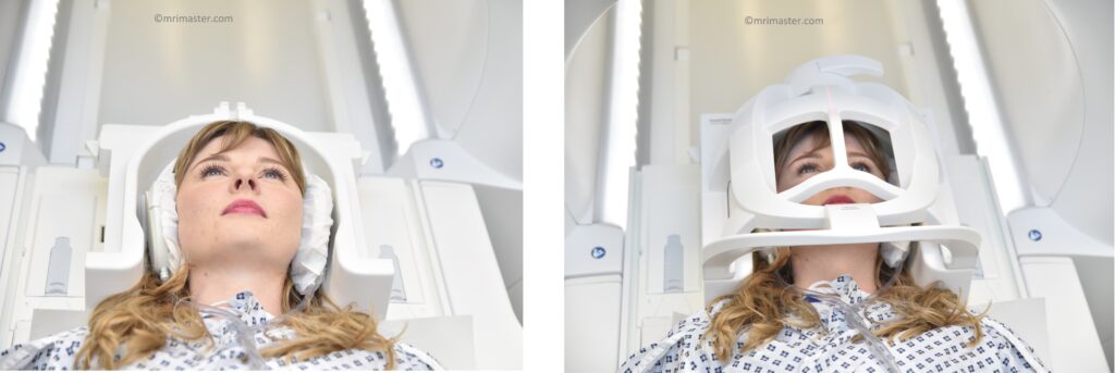
Positioning for Trigeminal nerve MRI
- Head first supine
- Position the head in the head coil and immobilise with cushions
- Give cushions under the legs for extra comfort
- Centre the laser beam localizer over the glabella
Recommended Trigeminal Nerve MRI Protocols and Planning
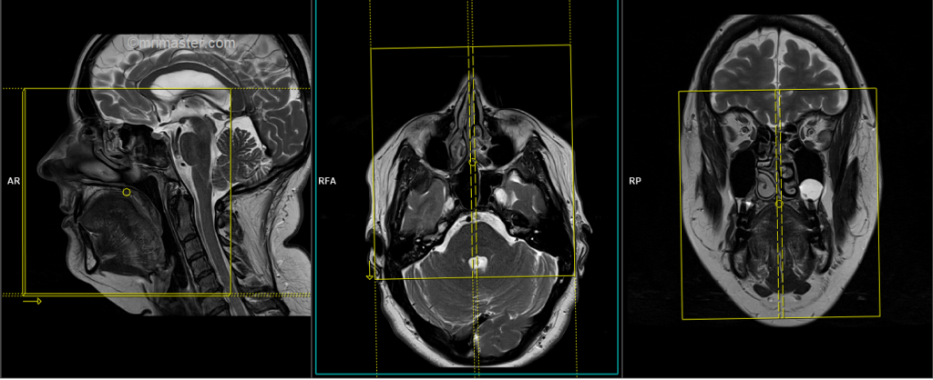
Trigeminal nerve MRI localiser
To plan the sequences, it is necessary to acquire a three-plane localizer, which typically consists of T1 weighted low-resolution scans lasting less than 25 seconds.

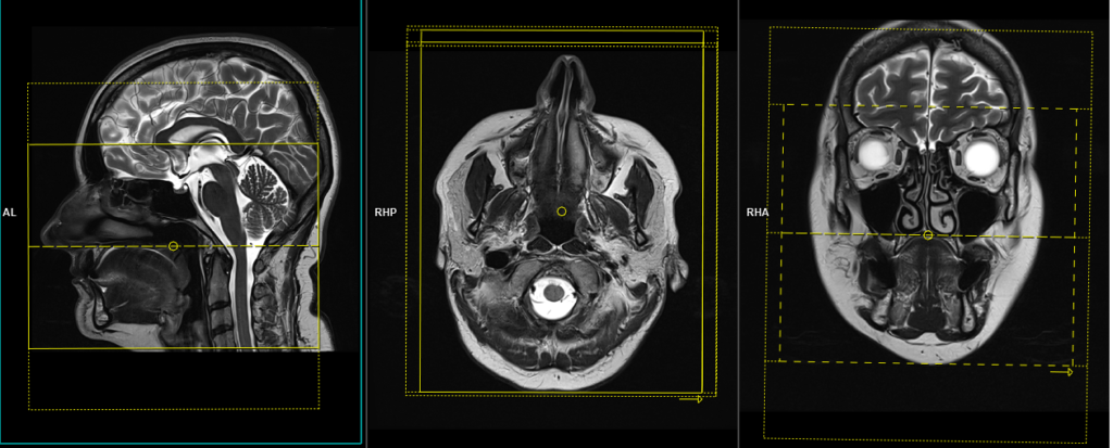
T2 STIR coronal 3mm SFOV
Plan the coronal slices on the sagittal plane; angle the positioning block perpendicular to the hard palate. Check the positioning block in the other two planes. Provide an appropriate angle in the axial plane (perpendicular to the nasal septum). The slices must be sufficient to cover the entire face from the nose to the level of the fourth ventricle.

Parameters
TR 5000-6000 | TE 110 | FLIP 130 | NEX 3 | SLICE 3 MM | MATRIX 256X256 | FOV 160-180 | PHASE R>L | GAP 10% | TI 150 |
T1 tse coronal 3mm small FOV
Plan the coronal slices on the sagittal plane; angle the positioning block perpendicular to the hard palate. Check the positioning block in the other two planes. Provide an appropriate angle in the axial plane (perpendicular to the nasal septum). The slices must be sufficient to cover the entire face from the nose to the level of the fourth ventricle.
Parameters
TR 400-600 | TE 15-25 | SLICE 3 MM | FLIP 140 | PHASE R>L | MATRIX 256X256 | FOV 160-180 | GAP 10% | NEX(AVRAGE) 2 |
T2 3D SPACE or CISS coronal .9mm
Plan the coronal 3D block on the sagittal plane; angle the positioning block perpendicular to the hard palate. Check the positioning block in the other two planes. An appropriate angle must be given in the axial plane (perpendicular to the nasal septum). Slices must be sufficient to cover the trigeminal main branches from the frontal slice to the level of the fourth ventricle.

Parameters
TR 2000-2500 | TE 250-300 | SLICE .9MM | FLIP 150 | PHASE R>L | MATRIX 320X320 | FOV 160-200 | GAP 10% | NEX(AVRAGE) 1.6 |
T2 tse axial 3mm SFOV
Plan the axial slices on the sagittal plane; angle the positioning block parallel to the hard palate. Check the positioning block in the other two planes. An appropriate angle must be given in the coronal plane (perpendicular to the nasal septum). Slices must be sufficient to cover the face from the glabella down to the angle of the jaw.

Parameters
TR 4000-5000 | TE 100-120 | SLICE 3 MM | FLIP 130-150 | PHASE R>L | MATRIX 288X256 | FOV 160-170 | GAP 10% | NEX(AVRAGE) 3 |
T2 tse sagittal 3mm SFOV
Plan the sagittal slices on the axial plane; angle the positioning block parallel to the nasal septum. Check the positioning block in the other two planes. An appropriate angle must be given in the coronal plane (parallel to the nasal septum). The slices should adequately cover the face from the right pinna to the left pinna.
Parameters
TR 4000-5000 | TE 100-120 | SLICE 3 MM | FLIP 130-150 | PHASE R>L | MATRIX 288X256 | FOV 160-170 | GAP 10% | NEX(AVRAGE) 3 |
For contrast enhanced scans
T1 VIBE DIXON axial .8mm SFOV ISO post contrast
Plan the axial 3D block on the sagittal plane and angle the positioning block parallel to the hard palate. Check the positioning block in the other two planes. An appropriate angle must be given in the coronal plane (perpendicular to the nasal septum). Slices should be sufficient to cover the face from the glabella down to the angle of the jaw.
Parameters
TR 7-8 | TE 2.39 4.77 | SLICE .8 MM | FLIP 12-15 | PHASE R>L | MATRIX 224X224 | FOV 200mm | GAP 10% | NEX(AVRAGE) 2 |