T1 SE / T1 TSE / T1 FSE Post Contrast
MRI image appearance
The easiest way to identify T1-weighted post-gadolinium images is by observing blood vessels in the body, such as arteries and veins in the brain, neck, chest, abdomen, upper limbs, and lower limbs. Blood vessels and pathologies with high vascularity appear bright on T1-weighted post-gadolinium images.
Tissues and their T1 post gadolinium appearance
Bone marrow : – equal to or higher than that of muscle (fatty marrow is usually bright)
Muscles- gray
Moving blood : – bright
White matter : – whiter
Gray matter : – gray
Fluids : – dark
Bone : – dark
Fat : – bright
Air : – dark
Use
- Very useful for brain and IAMs imaging
- Very useful for spine imaging
- Useful for pelvic imaging
- Useful for brachial and lumbar plexus imaging
- Useful for anterior neck orbits and face imaging
- Useful for any musculoskeletal imaging
- Useful for extremity imaging
Pathological appearance
Pathologies with hypervascularization will appear bright on T1-weighted post-gadolinium images. These include tumors like hemangioma, lymphangioma, hemangioendothelioma, Kaposi sarcoma, angiosarcoma, hemangioblastoma, as well as inflammatory processes like discitis, meningitis, synovitis, arthritis, and osteomyelitis. Pathological processes with no vascularity will remain unenhanced, appearing dark on T1-weighted post-gadolinium images.
* Most of the post contrast examinations use fat saturated post contrast T1 sequences except brain imaging.*
T1 AXIAL POST CONTRAST SEQUENCE USED IN BRAIN IMAGING

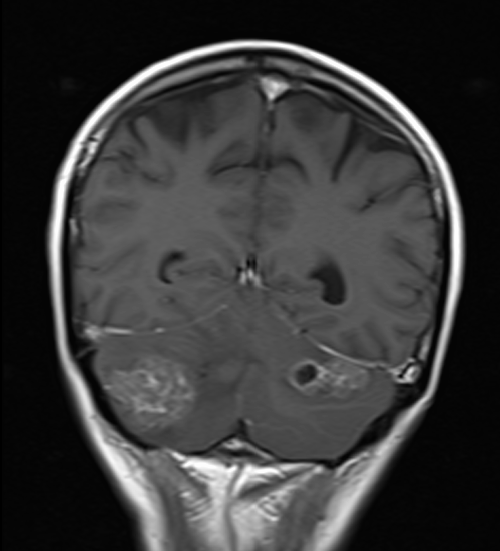
T1 CORONAL POST CONTRAST SEQUENCE USED IN BRAIN IMAGING
T1 CORONAL POST CONTRAST SEQUENCE USED IN IAMS IMAGING

T1 CORONAL POST CONTRAST SEQUENCE USED IN PNS IMAGING
