Abdominal Wall MRI
Indications for abdominal wall mri scan
- Abdominal wall endometriosis
- Abdominal wall muscle tear
- Abdominal wall infections
- Abdominal adhesions
- Abdominal wall fistula
- Abdominal wall masses
- Abdominal wall hernias
- Abdominal wall lipoma
Contraindications
- Any electrically, magnetically or mechanically activated implant (e.g. cardiac pacemaker, insulin pump biostimulator, neurostimulator, cochlear implant, and hearing aids)
- Intracranial aneurysm clips (unless made of titanium)
- Pregnancy (risk vs benefit ratio to be assessed)
- Ferromagnetic surgical clips or staples
- Metallic foreign body in the eye
- Metal shrapnel or bullet
Patient preparation for Abdominal Wall MRI
- A satisfactory written consent form must be taken from the patient before entering the scanner room
- Ask the patient to remove all metal objects including keys, coins, wallet, cards with magnetic strips, jewellery, hearing aid and hairpins
- Ask the patient to undress and change into a hospital gown
- Instruct the patient to hold their breath for the breath hold scans and breathe gently for the gated scans (its advisable to coach the patient two to three times before starting the scan)
- Buscopan injection risk and benefits must be explained to the patient before the scan
- Gadolinium should only be given to the patient if GFR is > 30
- An intravenous line must be placed with extension tubing extending out of the magnetic bore
- Claustrophobic patients may be accompanied into the scanner room e.g. by staff member or relative with proper safety screening
- Offer headphones for communicating with the patient and ear protection
- Explain the procedure to the patient and answer questions
- Note down the weight of the patient
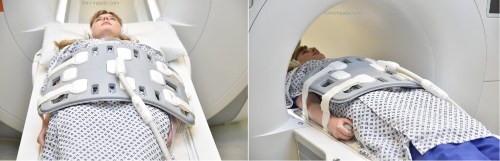
Positioning for Abdominal Wall MRI
- Position the patient in supine position with head pointing towards the magnet (head first supine).
- Position the patient over the spine coil and place the body coils over abdomen and pelvis (nipple down to three inches below symphysis pubis).
- Securely tighten the body coil using straps to prevent respiratory artefacts.
- Give a pillow under the head and cushions under the legs for extra comfort.
- Register the patient in the scanner as head first supine.
Recommended Abdominal Wall MRI Protocols and Planning
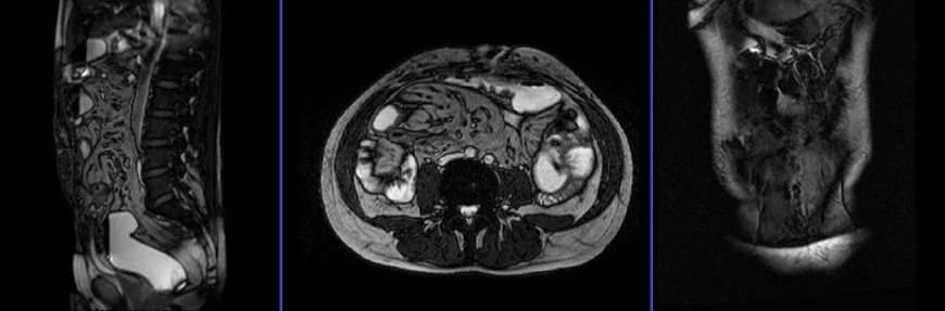
Abdominal Wall MRI localiser
A three-plane T2 TrueFISP/HASTE localizer must be taken initially to localize and plan the sequences. These are fast single-shot localizers with under 25s acquisition time, which are excellent for localizing abdominal structures.
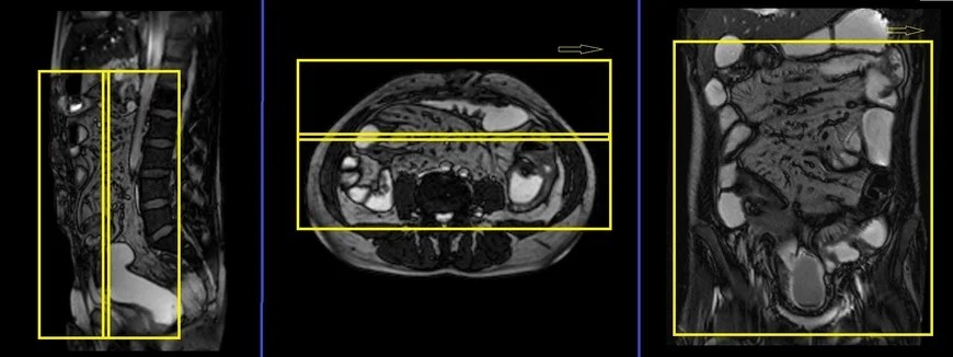
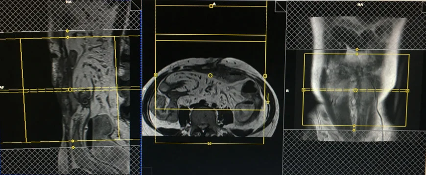
T2 TRUEFISP\HASTE coronal 4mm large FOV
Plan the coronal slices on the axial image, positioning the block horizontally across the abdomen as shown below. Verify the positioning block in the other two planes. Ensure an appropriate angle is set in the sagittal plane, vertically across the abdomen. The slices should adequately cover the entire abdomen from the anterior abdominal wall to the spinal canal. The field of view (FOV) must be large enough to encompass the abdomen and pelvis, from the stomach to the pubic symphysis. To prevent wrap-around artifacts, utilize phase oversampling. Instruct the patient to hold their breath during image acquisition.
Parameters TRUEFISP
TR 4-5 | TE 2-3 | FLIP 60 | NEX 1 | SLICE 4 MM | MATRIX 320×320 | FOV 400-450 | PHASE R>L | OVERSAMPLE 50% | IPAT OFF |
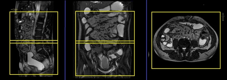
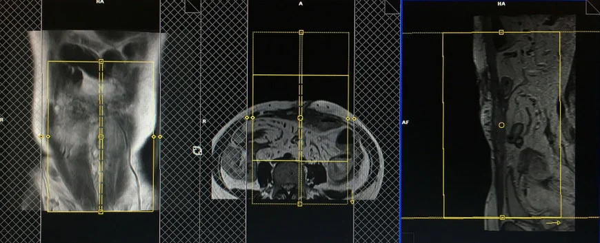
T2 TRUEFISP\HASTE axial 4MM Large FOV
Plan the axial slices on the coronal image, positioning the block horizontally across the abdomen as shown below. Check the positioning block in the other two planes. Ensure an appropriate angle is set in the sagittal plane, horizontally across the abdomen. Slices must be sufficient to cover the whole abdomen and pelvis from the stomach superiorly to the pubic symphysis inferiorly. Phase oversampling can be used to avoid wrap-around artifacts. Instruct the patient to hold their breath during image acquisition.
Parameters TRUEFISP
TR 4-5 | TE 2-3 | FLIP 60 | NEX 1 | SLICE 4 MM | MATRIX 320×256 | FOV 350-400 | PHASE A>P | OVERSAMPLE 50% | IPAT ON |
Pause for buscopan injection
Before the small field of view (SFOV) high-resolution scans, intravenously inject 0.5 to 1 ml (as per body weight and radiologist’s recommendation) of Buscopan. Wait for 1 minute before starting the next scan, as Buscopan needs time to take effect.
warning
* Buscopan injection should not be administered to patients with myasthenia gravis, megacolon, narrow-angle glaucoma, tachycardia, prostatic enlargement with urinary retention, mechanical stenoses in the region of the gastrointestinal tract, or paralytic ileus.*
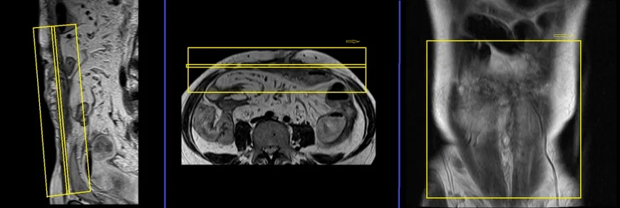
T2 TSE \HASTE axial multiple breath holds 4mm SFOV
Plan the axial slices on the sagittal image and angle the positioning block perpendicular to the rectus abdominis muscle. Verify the positioning block in the other two planes. Determine an appropriate angle in the coronal plane, perpendicular to the linea alba. Ensure that the slices are sufficient to cover the abdominal wall pathology. Choose a SFOV (Field of View) that is large enough to cover the affected area (typically 200mm-250mm). Utilize phase oversampling to avoid wrap-around artifacts. Additionally, add saturation bands on the top and bottom of the axial block to reduce arterial pulsation and breathing artifacts. Finally, instruct the patient to hold their breath during image acquisition.
This sequence is a variation of the T2 TSE (turbo spin echo) breath hold scan commonly used in abdominal and liver imaging. To achieve high-resolution breath hold scans, the user can make modifications to the T2 sequence. The default sequence parameters are as follows: field of view (FOV) of 350-400, matrix size of 256×256, number of excitations (NEX) set to 1, slice thickness of 6mm, and acquisition of 25-30 slices within a 30-second breath hold.
To attain the desired high resolution, the sequence can be adjusted to: FOV of 200-250, matrix size of 256×139, NEX of 2, and slice thickness of 4mm, with parallel imaging (IPAT) enabled. Consequently, the adapted sequence will take around 75 seconds, which should be divided into 3 acquisitions (concatenations). Each breath hold acquisition will be approximately 25 seconds.

Parameters T2 TSE
TR 2500-3000 | TE 80-100 | BW 170 | NEX 2 | SLICE 4 MM | MATRIX 256X192 | FOV 200-250 | PHASE A>P | OVERSAMPLE 50% | IPAT ON |
The T2 TSE images exhibited superior quality compared to the HASTE images. Nevertheless, in cases where the patient is unable to hold their breath, consider utilizing HASTE breath-hold or respiratory-gated HASTE sequences.
T1 VIBE DIXON SFOV 4mm axial breath hold
Plan the axial slices on the sagittal image and angle the positioning block perpendicular to the rectus abdominis muscle. Verify the positioning block in the other two planes. Determine an appropriate angle in the coronal plane, perpendicular to the linea alba. Ensure that the slices are sufficient to cover the abdominal wall pathology. Choose a SFOV (Field of View) that is large enough to cover the affected area (typically 200mm-250mm). Utilize phase oversampling to avoid wrap-around artifacts. Additionally, add saturation bands on the top and bottom of the axial block to reduce arterial pulsation and breathing artifacts. Finally, instruct the patient to hold their breath during image acquisition.
Parameters
TR 6-7 | TE 2.39 4.77 | FLIP 10 | NXA 1 | SLICE 3 MM | MATRIX 256×256 | FOV 200-250 | PHASE A>P | OVERSAMPLE 20% | BH YES |
T2 TSE SPAIR\HASTE FS axial multiple breath holds 4mm SFOV
Plan the axial slices on the sagittal image and angle the positioning block perpendicular to the rectus abdominis muscle. Verify the positioning block in the other two planes. Determine an appropriate angle in the coronal plane, perpendicular to the linea alba. Ensure that the slices are sufficient to cover the abdominal wall pathology. Choose a SFOV (Field of View) that is large enough to cover the affected area (typically 200mm-250mm). Utilize phase oversampling to avoid wrap-around artifacts. Additionally, add saturation bands on the top and bottom of the axial block to reduce arterial pulsation and breathing artifacts. Finally, instruct the patient to hold their breath during image acquisition.
Parameters
TR 3000-3500 | TE 80-100 | BW 150 | NEX 2 | SLICE 4 MM | MATRIX 224X192 | FOV 200-250 | PHASE A>P | OVERSAMPLE 50% | IPAT ON |
T2 TSE sagittal multiple breath holds 4mm SFOV
Plan the sagittal slices on the coronal image and angle the positioning block parallel to the linea alba. Verify the positioning block in the other two planes. Provide an appropriate angle in the axial plane perpendicular to the rectus abdominis muscle. The slices must be sufficient to cover the abdominal wall pathology. Choose a small field of view (FOV) that is sufficient to cover the affected area (normally 200mm-250mm). Phase oversampling must be used to avoid wrap-around artifacts. Instruct the patient to hold their breath during image acquisition.
Parameters
TR 2500-3000 | TE 80-100 | BW 170 | NEX 2 | SLICE 4 MM | MATRIX 256X192 | FOV 200-250 | PHASE A>P | OVERSAMPLE 50% | IPAT ON |
T1 VIBE DIXON SFOV 4mm sagittal breath hold
Plan the sagittal slices on the coronal image and angle the positioning block parallel to the linea alba. Verify the positioning block in the other two planes. Provide an appropriate angle in the axial plane perpendicular to the rectus abdominis muscle. The slices must be sufficient to cover the abdominal wall pathology. Choose a small field of view (FOV) that is sufficient to cover the affected area (normally 200mm-250mm). Phase oversampling must be used to avoid wrap-around artifacts. Instruct the patient to hold their breath during image acquisition.

Parameters
TR 6-7 | TE 2.39 4.77 | FLIP 10 | NXA 1 | SLICE 3 MM | MATRIX 256×256 | FOV 200-250 | PHASE A>P | OVERSAMPLE 20% | BH YES |
T2 TSE\HASTE coronal multiple breath holds 4mm SFOV
Plan the coronal slices on the sagittal image, angling the positioning block parallel to the rectus abdominis muscle. Verify the positioning block in the other two planes. Provide an appropriate angle in the axial plane, horizontally across the rectus abdominis muscle. The slices must be sufficient to cover the abdominal wall pathology. Choose a small field of view (FOV) that is sufficient to cover the affected area (normally 200mm-250mm). Phase oversampling must be used to avoid wrap-around artifacts. Instruct the patient to hold their breath during image acquisition.
Parameters
TR 2500-3000 | TE 80-100 | BW 170 | NEX 2 | SLICE 4 MM | MATRIX 256X192 | FOV 200-250 | PHASE R>L | OVERSAMPLE 50% | IPAT ON |