MRI Forefoot : Protocol and planning
Indications Forfoot MRI scan
- Marrow abnormalities (eg. bone contusions, osteonecrosis, marrow edema syndromes, and stress fractures)
- Synovial based disorders ( e.g. synovitis, tenosynovitis, bursitis, and ganglion cysts)
- Congenital and developmental conditions ( eg.dysplasia, tarsal coalition)
- Infections of bone, joint or soft tissue (e.g. osteomyelitis ,osteoarthritis )
- Plantar fasciitis, fascial rupture and plantar fibromatosis
- Neoplasms of bone, joint or soft tissue
- Abnormalities of other hindfoot tendons
- Abnormalities of foot tendons
- Mortons neuroma
- Avascular necrosis
Contraindications
- Any electrically, magnetically or mechanically activated implant (e.g. cardiac pacemaker, insulin pump biostimulator, neurostimulator, cochlear implant, and hearing aids)
- Intracranial aneurysm clips (unless made of titanium)
- Pregnancy (risk vs benefit ratio to be assessed)
- Ferromagnetic surgical clips or staples
- Metallic foreign body in the eye
- Metal shrapnel or bullet
Patient preparation for Forfoot MRI scan
- A satisfactory written consent form must be taken from the patient before entering the scanner room
- Ask the patient to remove all metal object including keys, coins, wallet, any cards with magnetic strips, jewellery, hearing aid and hairpins
- If possible provide a chaperone for claustrophobic patients (e.g. relative or staff )
- Offer earplugs or headphones, possibly with music for extra comfort
- Explain the procedure to the patient
- Instruct the patient to keep still
- Note the weight of the patient
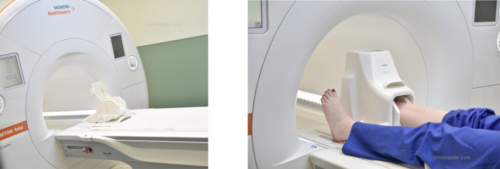
Positioning for Forfoot MRI scan
- Position the patient in supine position with feet pointing towards the magnet (feet first supine)
- Position the ankle over the foot and ankle coil (use head coil if ankle coil is not available) and lock it properly (foot should be flexed 90° and flatten to get good scans)
- Securely tighten the foot using cushions to prevent movement
- Give a pillow under the head for extra comfort
- Centre the laser beam localiser over foot
Recommended Forfoot MRI Protocols and Planning
localiser
A three-plane localizer must be taken at the beginning to localize and plan the sequences. Localizers are usually less than 25 seconds. T1-weighted low-resolution scans.(plan a second localiser if needed).

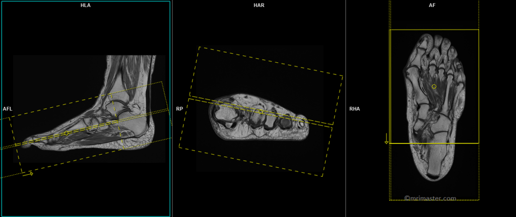
T1 tse axial 3 mm (coronal in anatomical position)
Plan the axial slices on the sagittal plane, angling the positioning block perpendicular to the metatarsal and phalanges bones. Check the positioning block in the other two planes. An appropriate angle must be given in the coronal plane (perpendicular to the metatarsal and phalanges bones). The slices must be sufficient to cover the forefoot from the tip of the toe up to the metatarsal bones. Most of the radiologists refer this as an axial plane even though in anatomical position this is coronal plane

Parameters
TR 500-600 | TE 15-25 | SLICE 3 MM | FLIP 90 | PHASE A>P | MATRIX 304X256 | FOV 120-140 | GAP 10% | NEX(AVRAGE) 2 |
T2 stir axial 3 mm
Plan the axial slices on the sagittal plane, angling the positioning block perpendicular to the metatarsal and phalanges bones. Check the positioning block in the other two planes. An appropriate angle must be given in the coronal plane (perpendicular to the metatarsal and phalanges bones). The slices must be sufficient to cover the forefoot from the tip of the toe up to the metatarsal bones.
Parameters
TR 4000-5000 | TE 110 | FLIP 150 | NEX 3 | SLICE 3 MM | MATRIX 256X256 | FOV 120-140 | PHASE A>P | GAP 10% | TI 150 |
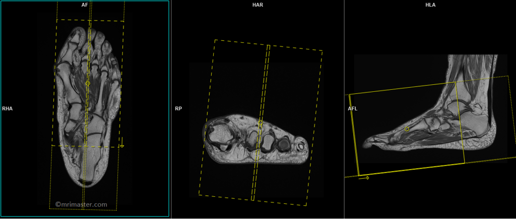
T1 tse coronal 2mm
Plan the coronal slices on the sagittal plane, ensuring that the positioning block is angled parallel to the metatarsal and phalanges bones. Verify the positioning of the block in the other two planes. Establish an appropriate angle in the axial plane, aligning it parallel to the line across the metatarsal bones. The slices should adequately cover the foot from the plantar aspect up to the dorsal aspect. Use a field of view (FOV) small enough, such as 120 to 150mm, to encompass the metatarsal and phalanges bones. Most of the radiologists refer this as a coronal plane even though in anatomical position this is an axial plane.
Parameters
TR 400-600 | TE 15-25 | SLICE 2 MM | NEX 2 | PHASE A>P | MATRIX 304X304 | FOV 120-150 | GAP 10% | OVERSAMPLE 70% |
T2 stir coronal 2mm
Plan the coronal slices on the sagittal plane, ensuring that the positioning block is angled parallel to the metatarsal and phalanges bones. Verify the positioning of the block in the other two planes. Establish an appropriate angle in the axial plane, aligning it parallel to the line across the metatarsal bones. The slices should adequately cover the foot from the plantar aspect up to the dorsal aspect. Use a field of view (FOV) small enough, such as 120 to 150mm, to encompass the metatarsal and phalanges bones. Most of the radiologists refer this as a coronal plane even though in anatomical position this is an axial plane.
Parameters
TR 4000-5000 | TE 110 | OS 70% | NEX 3 | SLICE 2 MM | MATRIX 304X256 | FOV 120-150 | PHASE A>P | GAP 10% | TI 150 |
T1 tse sagittal 2mm
Plan the sagittal slices on the coronal plane and angle the positioning block parallel to the metatarsal and phalanges bones. Verify the block’s positioning in the other two planes. Establish an appropriate angle in the axial plane, perpendicular to the metatarsal bones. Ensure that the slices adequately cover the foot from side to side. Utilize a small FOV (e.g., 120 to 150mm) to encompass the metatarsal and phalanges bones.
Parameters
TR 400-600 | TE 15-25 | SLICE 2 MM | NEX 2 | PHASE A>P | MATRIX 320X320 | FOV 120-150 | GAP 10% | OVERSAMPLE 70% |
T2 stir sagittal 2mm
Plan the sagittal slices on the coronal plane and angle the positioning block parallel to the metatarsal and phalanges bones. Verify the block’s positioning in the other two planes. Establish an appropriate angle in the axial plane, perpendicular to the metatarsal bones. Ensure that the slices adequately cover the foot from side to side. Utilize a small FOV (e.g., 120 to 150mm) to encompass the metatarsal and phalanges bones.
Parameters
TR 4000-5000 | TE 110 | OS 70% | NEX 3 | SLICE 2 MM | MATRIX 256X256 | FOV 120-150 | PHASE A>P | GAP 10% | TI 150 |