Aliasing / Wrap around artifact
Aliasing, also known as wrap-around artifact, is a common artifact that can occur in magnetic resonance imaging (MRI) when the field of view (FOV) is smaller than the actual object being imaged. Spatial localisation is determined by the frequency of the spins within a selected sample of tissue. Spins within a chosen FOV have a phase shift of 0-360°. If the digital sampling rate is too low (less than twice the highest frequency contained within that signal), spins outside the FOV with a phase shift of less than 0 or over 360° may be encoded along with spins inside the FOV that have the same phase value. eg. a spin with shift of 365° will be mis-mapped to the same position as a spin of 5° and appear on the other side of the image.
In 3D imaging phase encoding is used to select sections of tissue. If the anatomy extends beyond the FOV, a similar process may cause wrap-over in the slab select direction between slices at either end of the volume.

Here are some strategies to minimize or avoid Aliasing Artifacts
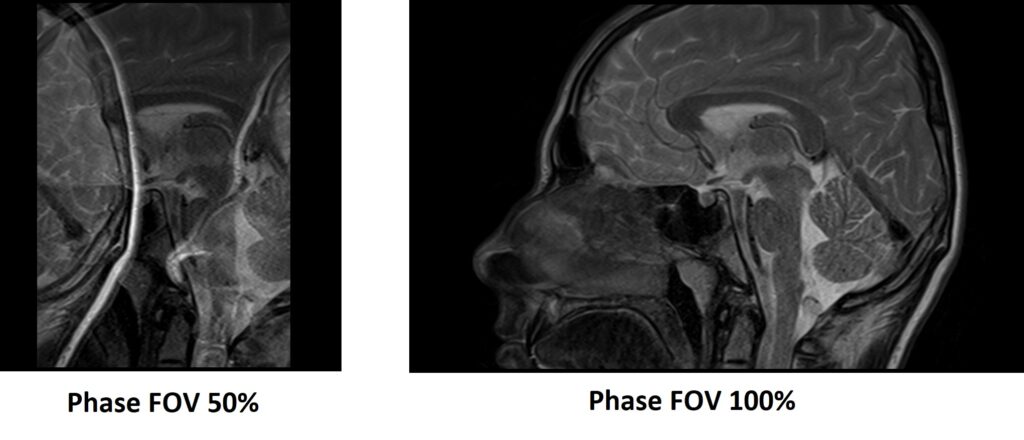
Increase field of view (FOV): One way to minimize aliasing artifacts is by increasing the field of view (FOV). This can be accomplished by expanding either the imaging FOV or the FOV in the phase-encoding direction.
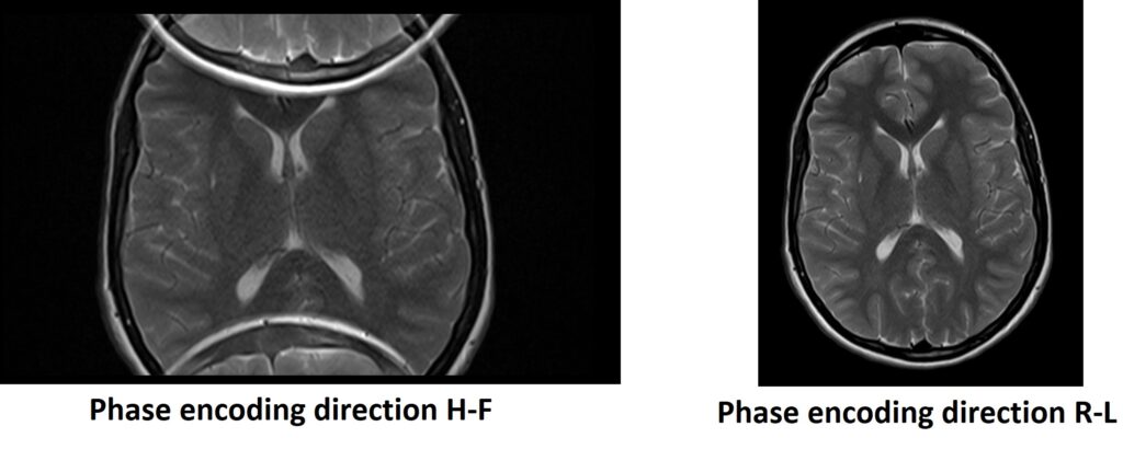
Adjust the phase-encoding direction: Altering the phase-encoding direction can help mitigate aliasing artifacts. Choosing a phase-encoding direction that is less susceptible to aliasing, such as selecting the superior-inferior direction instead of the anterior-posterior or left-right direction, can be beneficial.
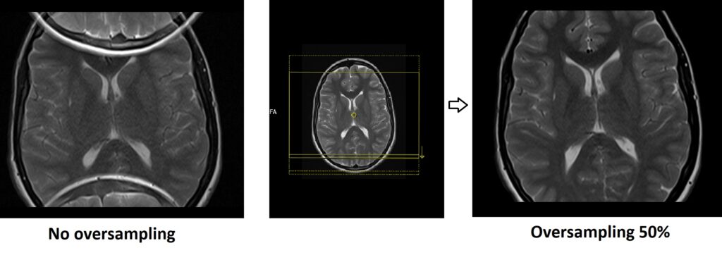
Oversampling in the phase-encoding direction: Acquiring additional phase-encoding steps beyond what is necessary for the desired FOV can help reduce aliasing artifacts. This oversampling allows for a better estimation and reconstruction of the image without aliasing.
Place saturation bands outside FOV in the phase-encoding (PE) direction: Saturation bands are radiofrequency pulses applied to selectively saturate the signal from tissues outside the region of interest. By placing saturation bands outside the FOV in the PE direction, you can effectively suppress the signal from those regions, reducing the likelihood of wrap-around artifacts.
Use surface coils: Surface coils are smaller and positioned closer to the area of interest. They provide improved spatial resolution and reduced sensitivity to signals from outside the region of interest. By using surface coils instead of larger volume coils, you can minimize the signal contribution from tissues outside the desired imaging area, thereby reducing the chances of wrap-around artifacts.
References
- Hu, R., Liu, Y., & Li, X. (2021). Simulation and Mitigation of the Wrap-Around Artifact in the MRI Image. Front Comput Neurosci, 15, 746549. doi: 10.3389/fncom.2021.746549. PMID: 34744675. PMCID: PMC8566355.
- Pusey, E., Yoon, C., Anselmo, M. L., & Lufkin, R. B. (1988). Aliasing artifacts in MR imaging. Comput Med Imaging Graph, 12(4), 219-224. doi: 10.1016/0895-6111(86)90003-0. PMID: 3179974.
- Runge, V. M., & Price, A. C. (2015). Magnetic Resonance Imaging: Physical Principles and Sequence Design. John Wiley & Sons.