Contrast-to-Noise Ratio (CNR) in MRI
CNR stands for Contrast-to-Noise Ratio in MRI. It is a metric used to assess the quality of an MRI image by measuring the contrast between different tissues or regions of interest (ROIs) in relation to the background noise. A higher CNR indicates better image quality, as it signifies a clearer distinction between the areas being compared.
Here’s a more detailed breakdown:
Contrast: This refers to the difference in signal intensity between two areas in the image. For example, it could be the difference between a tumor and the surrounding healthy tissue.
Noise: This is the random variation in the signal that is not due to the actual signal from the tissue. Noise can come from various sources, including the MRI machine itself, the environment, and the patient’s movements.
Ratio: The CNR is the ratio of the contrast (difference in signal intensities) to the noise level. It can be expressed mathematically as:
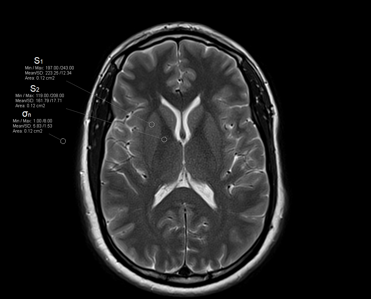
CNR = (S1 - S2) / σnwhere S1 and S2 are the signal intensities of the two regions being compared, and σn is the standard deviation of the noise.
A higher CNR is generally desirable as it implies that the differences between tissues or structures are more easily discernible against the background noise, leading to more precise and accurate diagnoses.
Steps to Calculate CNR in an MRI Image
Select Regions of Interest:
- Choose a region of interest (ROI) within the tissue or structure you are analyzing.
- Choose a background region or another tissue for comparison.
Measure Signal Intensities:
- Calculate the mean signal intensity (
S1) within the ROI. - Calculate the mean signal intensity (
S2) within the background or comparison region.
Measure Noise:
- Identify a region in the image that represents pure noise (usually in the background where there is no signal).
- Calculate the standard deviation of the signal intensities in this noise region (
σnoise).
Compute CNR:
- Use the formula:
CNR = (S1 - S2) / σnoiseto compute the CNR.
Example
Suppose in an MRI image:
- The mean signal intensity of the ROI (
S1) is 150 units. - The mean signal intensity of the background (
S2) is 50 units. - The standard deviation of the noise (
σnoise) is 10 units.
Then,
CNR = (150 - 50) / 10 = 100 / 10 = 10

Contrast-to-Noise Ratio (CNR) calculator for MRI
| Parameter | Symbol | Value |
|---|---|---|
| Signal Intensity of Tissue 1 | S1 | |
| Signal Intensity of Tissue 2 (or Background) | S2 | |
| Standard Deviation of Noise | σ |
Factors affecting CNR
Magnetic Field Strength
- Higher Field Strength: Increases the signal-to-noise ratio (SNR), which can improve CNR.
- Lower Field Strength: Reduces SNR, potentially decreasing CNR.
Pulse Sequence Parameters
- Repetition Time (TR): Longer TR can increase signal intensity, improving CNR for certain tissues.
- Echo Time (TE): Shorter TE minimizes signal decay, which can help in maintaining higher signal intensities and thus better CNR.
- Flip Angle: Optimizing the flip angle can maximize signal intensity, impacting CNR.
Receiver Bandwidth
- Narrower Bandwidth: Reduces noise, improving SNR and thus CNR.
- Wider Bandwidth: Increases noise, potentially reducing CNR.
Voxel Size
- Larger Voxel Size: Increases signal by capturing more signal from a larger volume, improving SNR and potentially CNR.
- Smaller Voxel Size: Reduces signal, decreasing SNR and potentially CNR.
Number of Signal Averages (NSA) or Number of Excitations (NEX)
- Higher NSA/NEX: Averaging multiple acquisitions reduces noise, improving SNR and thus CNR.
- Lower NSA/NEX: Increases noise, potentially reducing CNR.
Use of Contrast Agents
- Contrast Agents: Enhance the difference in signal intensity between different tissues, significantly improving CNR.
Coil Selection and Positioning
- Appropriate Coil: Using a coil suited to the region being imaged can improve SNR and CNR.
- Positioning: Properly positioning the coil relative to the area of interest can maximize signal capture and improve CNR.
Patient Factors
- Motion: Patient motion can blur images, reducing CNR. Techniques like motion correction and patient instructions to remain still can help.
- Body Size: Larger patients may have lower SNR due to increased distance between the coil and the area of interest, affecting CNR.
Imaging Environment
Magnetic Field Homogeneity: Uniform magnetic fields provide more consistent signal intensities, improving CNR.