CISS/FIESTA-C/Phase Balanced SARGE, PBSG MRI
CISS (Constructive Interference in Steady State) MRI is an advanced imaging technique derived from the TrueFISP sequence. It utilizes specialized radiofrequency pulses, gradient echo sequences, and a T2-weighted 3D gradient echo technique to produce high-resolution isotropic images. By executing two consecutive runs of 3D balanced steady-state free precession with varying excitation levels and then combining them, CISS accentuates the contrast between fluids, such as cerebrospinal fluid (CSF), and surrounding structures. This exceptional sensitivity to fluids enables precise visualization of intricate structures within the nervous system, including the brain, spinal cord, and inner ear. Its ability to provide heavily T2-weighted images sets it apart as a valuable tool for detailing soft tissues and distinguishing fine structures.
CISS MRI Physics
CISS is a gradient echo sequence that capitalizes on both gradient refocused and RF-refocused echoes, resulting in fluids exhibiting high signal intensity. Its operation involves two consecutive runs of 3D balanced steady-state free precession with varied excitation levels, which are combined subsequently. The image contrast within CISS is guided by the tissue’s T2/T1 ratio. Tissues displaying long T2 and short T1 relaxation times produce high signal intensity in CISS images, a feature seen prominently in both water and fat.
CISS MRI Image Appearance
In CISS imaging, cerebrospinal fluid (CSF) stands out brilliantly against surrounding tissues, offering exceptional contrast and resolution. This technique excels in depicting intricate structures like those in the posterior fossa, inner ear, and cranial nerves, as well as the spinal canal. Recognizing CISS images is straightforward—fluid-filled spaces, such as brain ventricles and spinal canals, appear exceptionally bright, while fat exhibits a bright but slightly less intense appearance.
CISS MRI Applications
Inner Ear: CISS MRI is often used to visualize the inner ear structures, including the cochlea, vestibule, and semicircular canals. It is particularly valuable in evaluating patients for potential causes of sensorineural hearing loss or for preoperative planning prior to cochlear implantation.
Cranial Nerves: The high resolution and contrast provided by CISS make it an excellent choice for imaging small structures like cranial nerves, especially in the cerebellopontine angle.
Spinal Imaging: CISS can be used to identify syrinx, CSF leak, spinal arteriovenous malformation, or cystic lesions in the spine.
CISS MRI Limitations
Prone to motion and flow artifacts.
Can present susceptibility artifacts in proximity to bone or air, resulting in signal voids.
Tissues and their CISS MRI appearance
Brain:
- CSF: Very bright.
- Fat: Bright but less intense than fluids.
- White Matter: Dark.
- Gray Matter: Dark.
- Bone (skull): Dark.
- Bone Marrow: Intermediate to bright.
- Salivary glands: Dark.
- Salivary duct: Very bright.
- Blood Vessels: Mostly Dark.
- Pituitary Gland: Dark.
- Choroid Plexus: Dark.
- Cerebellum: Dark.
- Brain Stem: Dark.
- Sinuses: Dark (air-filled).
- Thalamus, Putamen, Hippocampus, Caudate Nucleus: Dark.
- Corpus Callosum: Dark.
- Pineal Gland: Dark.
Spine:
- Spinal Cord: Dark.
- CSF: Very bright.
- Fat: Bright but less intense than fluids.
- Bone: Dark.
- Bone Marrow: Intermediate to bright.
- Intervertebral Disc: Intermediate.
- Ligaments: Intermediate to Dark.
- Nerve Roots: Dark.
Use
- Very useful for Cranial nerves imaging
- Useful for sailography imaging
- Useful for spinal cord imaging
- Useful for CSF leak imaging
- Useful for spinal AVM imaging
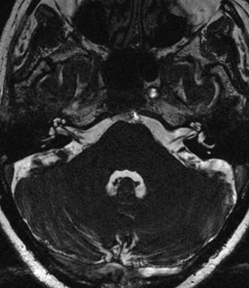
Axial CISS sequence used in IAMs imaging
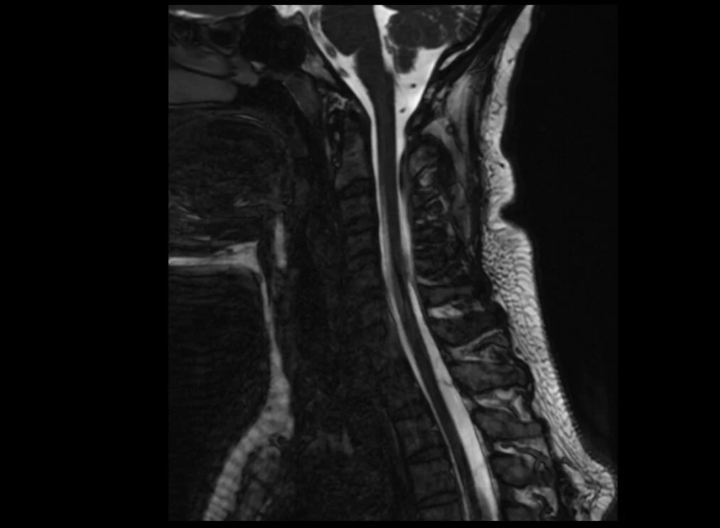
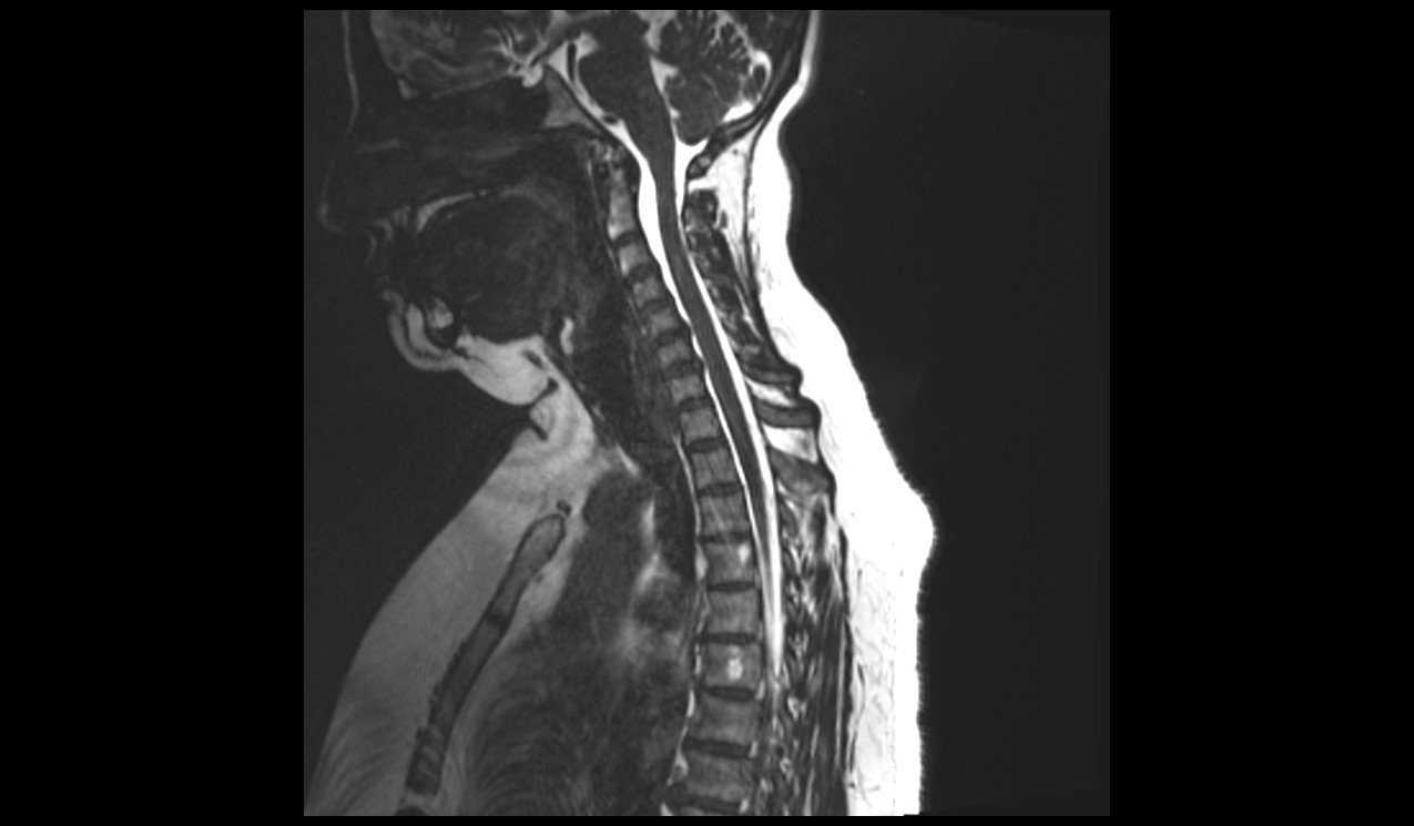
Sagittal CISS 3D sequence used in cervical spine imaging
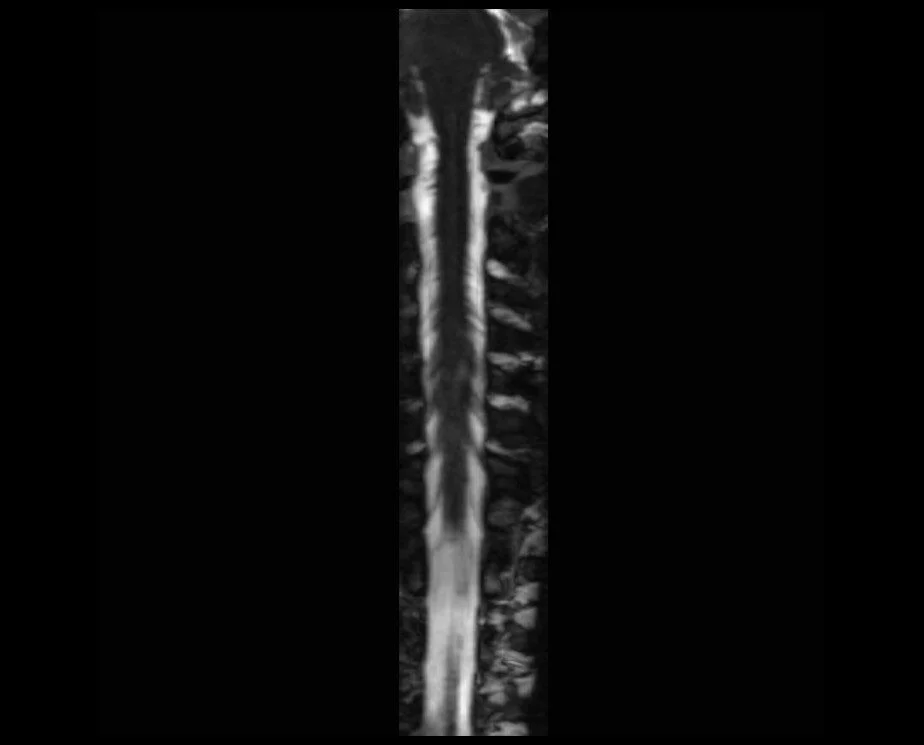
CISS 3D coronal reconstruction of cervical spinal cord
CISS 3D Axial reconstruction of cervical spinal cord

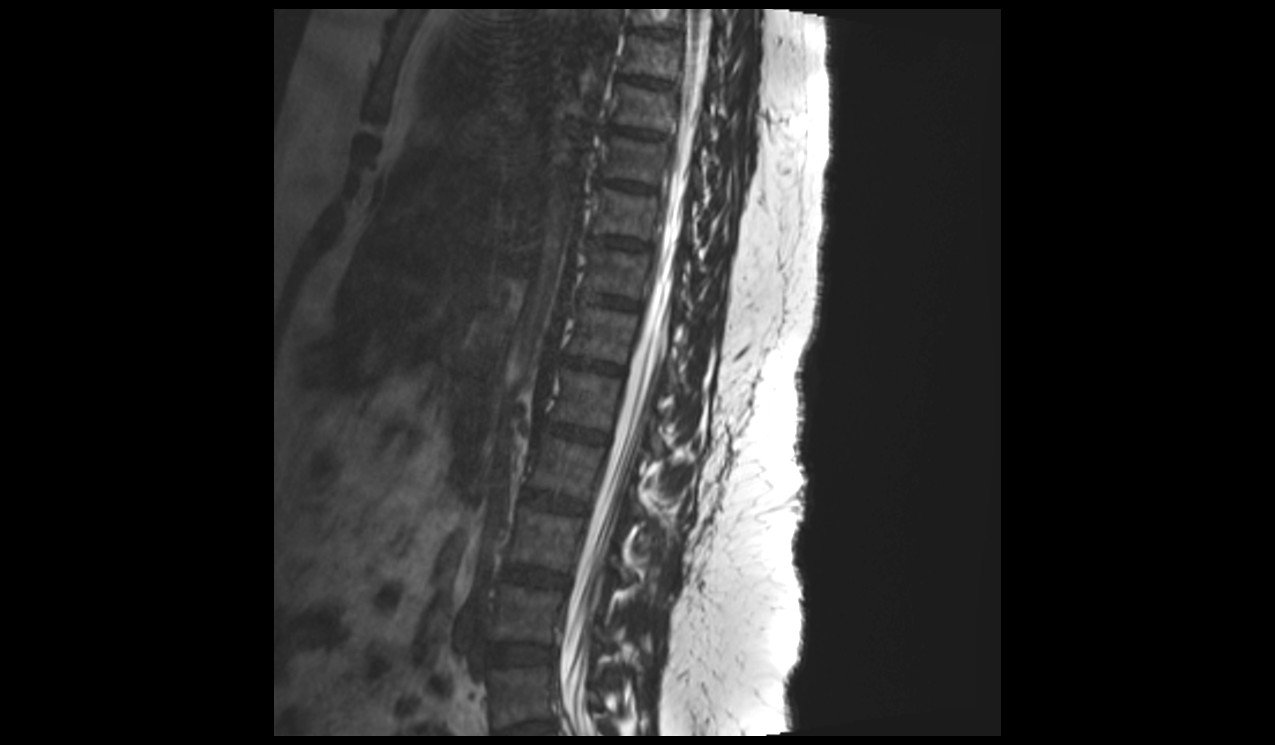
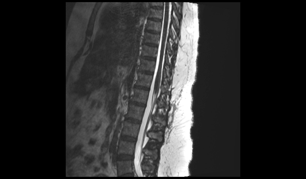
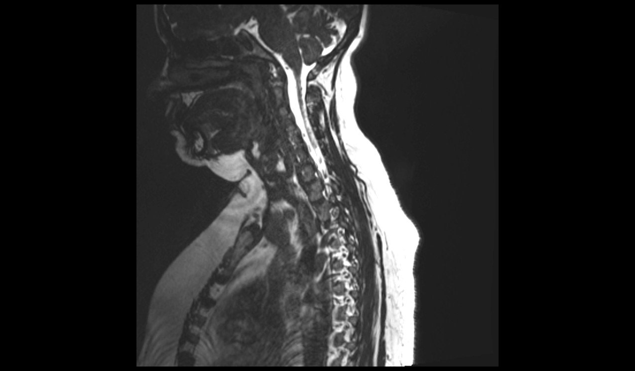
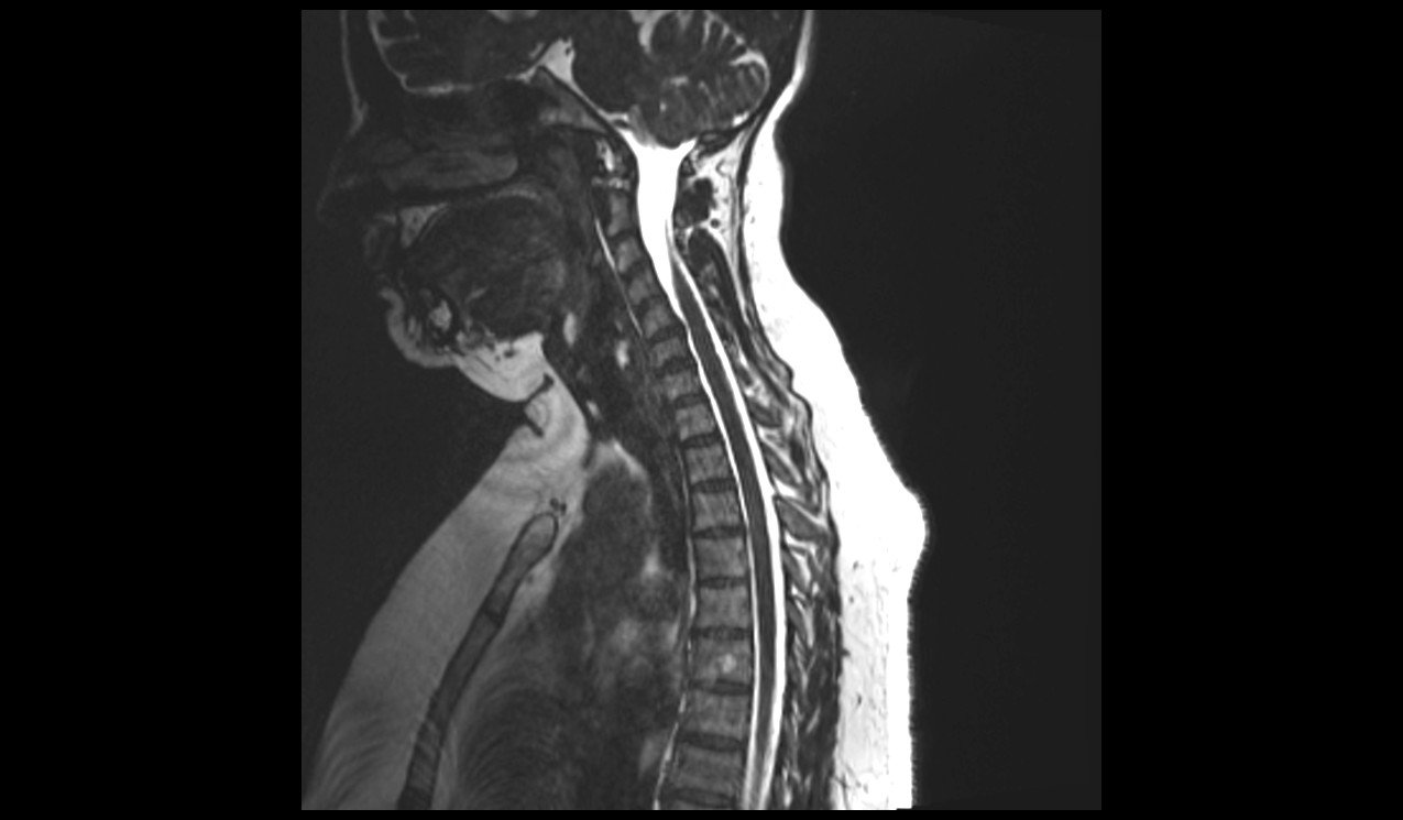
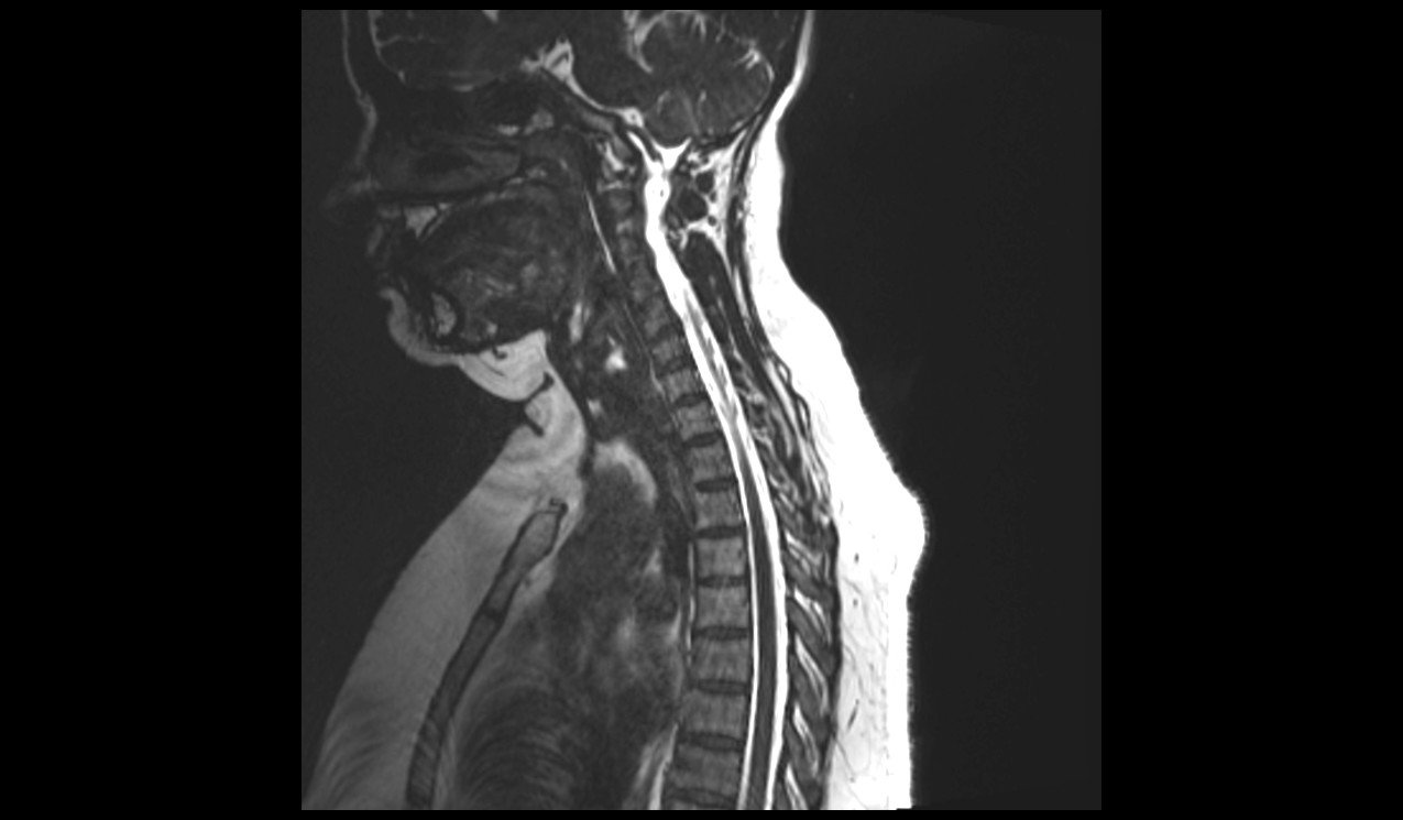
Sagittal CISS 3D sequence used in cervicothoracic spine imaging
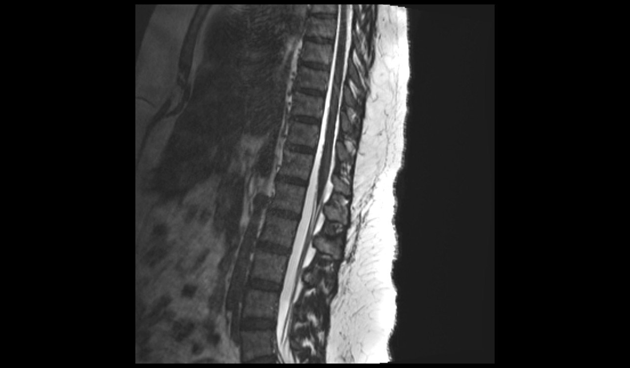
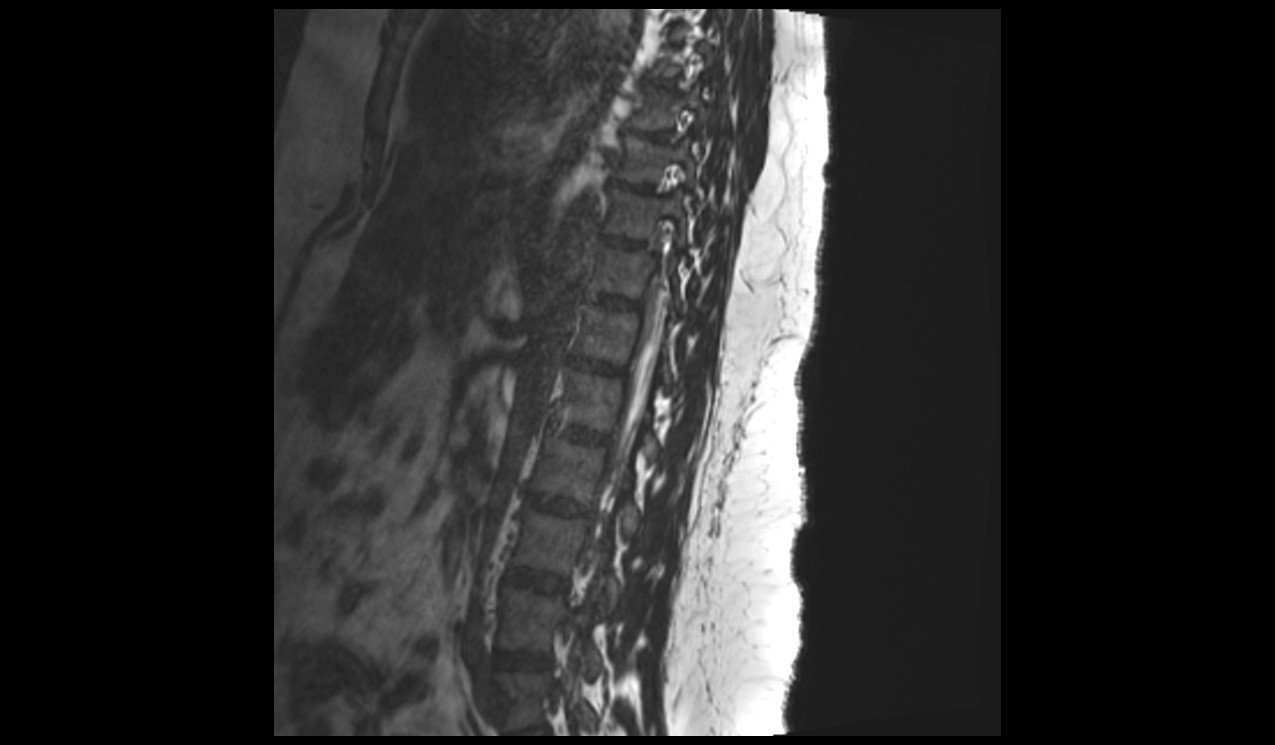
Sagittal CISS 3D sequence used in thoracolumbar spine imaging