RESOLVE DWI: Physics and Applications
RESOLVE (Readout SEgmentation Of Long Variable Echo-trains) DWI (Diffusion Weighted Imaging) is an advanced MRI (Magnetic Resonance Imaging) technique developed to obtain high-resolution diffusion-weighted images with reduced susceptibility-related artifacts. DWI is a powerful imaging method used to visualize the random motion of water molecules in tissues, offering insights into their microstructure. The RESOLVE technique further refines this method to offer clearer images, especially in anatomical regions that are prone to artifacts.
Physics of RESOLVE DWI
The fundamental principle behind DWI is based on the diffusion of water molecules. In standard DWI, the diffusion process is probed using a pair of magnetic field gradient pulses. The phase shifts caused by the motion of water molecules during the application of these pulses result in signal attenuation, which can be mathematically modeled and interpreted to produce an image.
However, in certain regions of the body, such as the base of the skull or near air-filled sinuses, susceptibility differences can result in magnetic field inhomogeneities. These lead to distortions and signal dropouts in echo planar imaging (EPI)-based DWI sequences.
RESOLVE DWI addresses this issue by segmenting the readout into multiple shorter echo trains. Instead of acquiring all k-space lines in a single shot (as in conventional single-shot EPI), RESOLVE splits the acquisition into several shots, each with fewer k-space lines. This reduces the echo spacing and the echo train length, thus minimizing the susceptibility-induced distortions.
Furthermore, RESOLVE incorporates a 2D navigator-based reacquisition scheme. This allows for the detection of shot-to-shot phase variations (caused by patient motion or system instabilities) and the subsequent correction or reacquisition of corrupted shots.
RESOLVE DWI Applications
- Neuroimaging: RESOLVE DWI significantly reduces susceptibility artifacts at the base of the brain, allowing for clearer visualization of regions such as the brainstem, temporal lobe, and cerebellum.
- Head & Neck Imaging: The intricate anatomy and proximity to air-filled sinuses make this region prone to susceptibility artifacts. RESOLVE offers superior visualization of these areas.
- Spinal Imaging: The vertebral column and its complex surrounding anatomy can be better assessed with RESOLVE, especially for conditions like spinal cord tumors or infections.
- Prostate Imaging: RESOLVE DWI offers high-resolution, artifact-free images of the prostate gland, enabling early detection of prostate cancer.
- Breast Imaging: DWI is increasingly used in breast imaging to differentiate benign from malignant tumors. RESOLVE further improves the clarity and detail of these images.
- Liver Imaging: RESOLVE aids in the detection of liver lesions, differentiation of benign from malignant tumors, and assessment of cirrhotic livers.
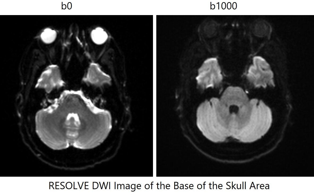
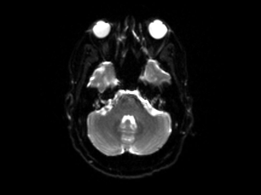
RESOLVE DWI Axial b0 Image of Brain
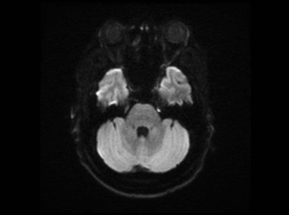
RESOLVE DWI Axial b1000 Image of Brain
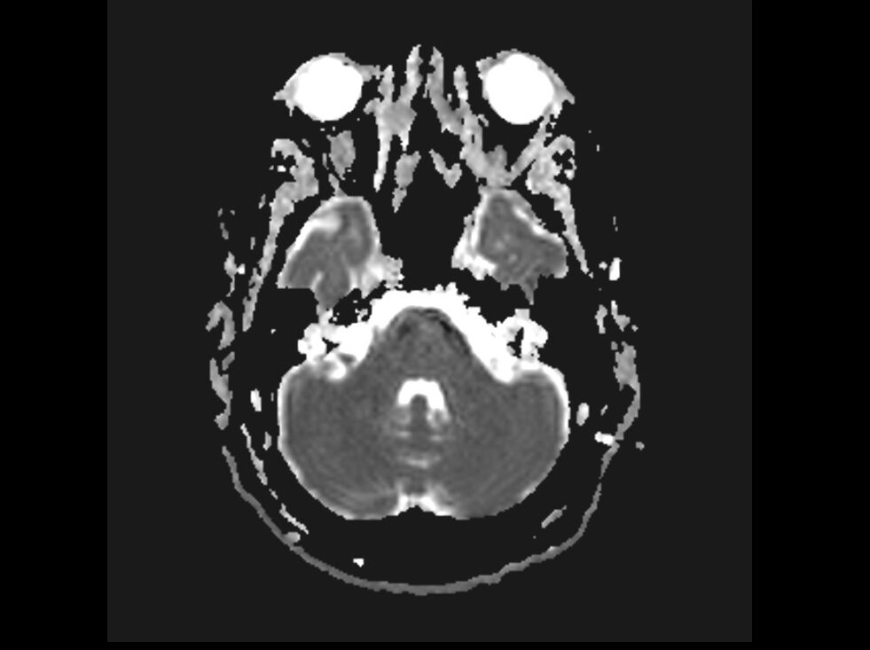
RESOLVE DWI Axial ADC Image of Brain
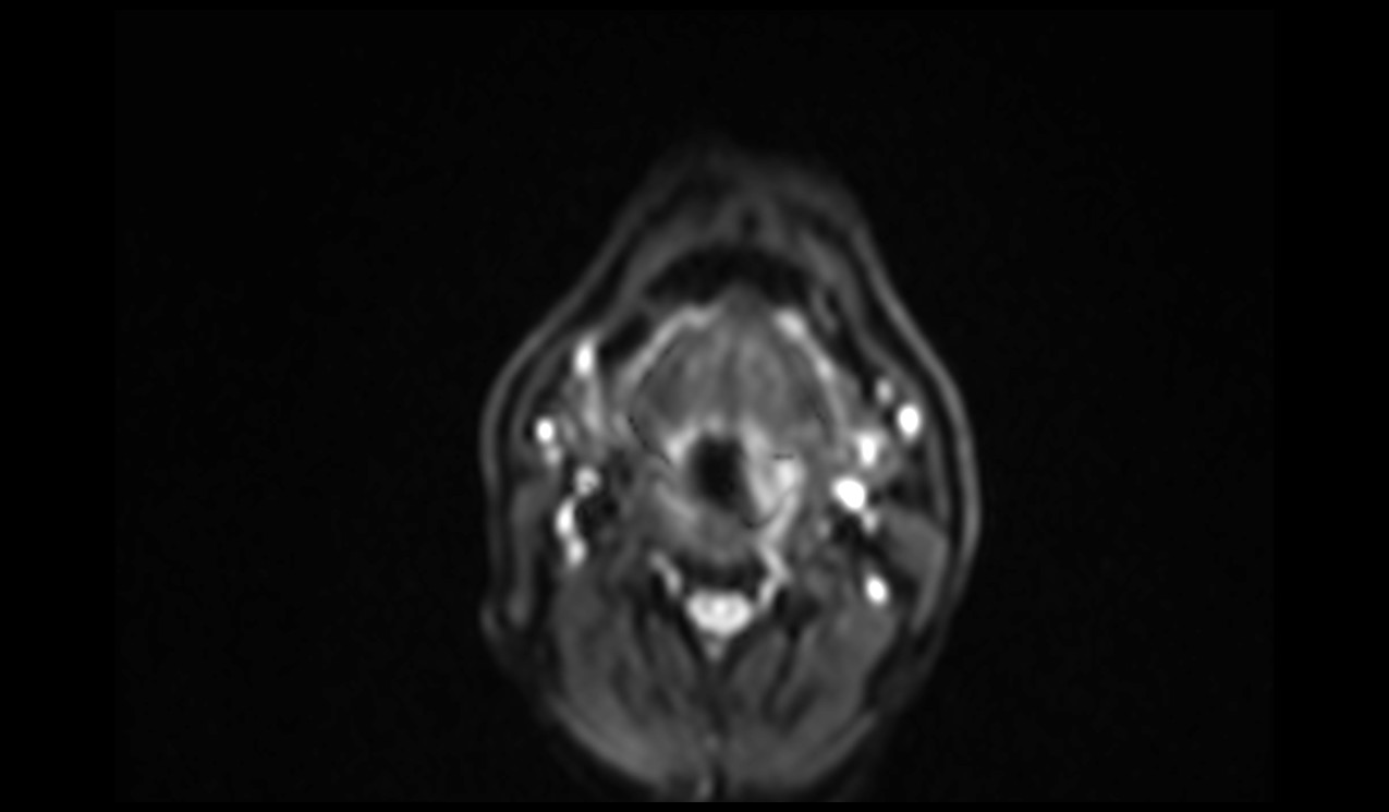
RESOLVE DWI Axial b50 Image of Neck
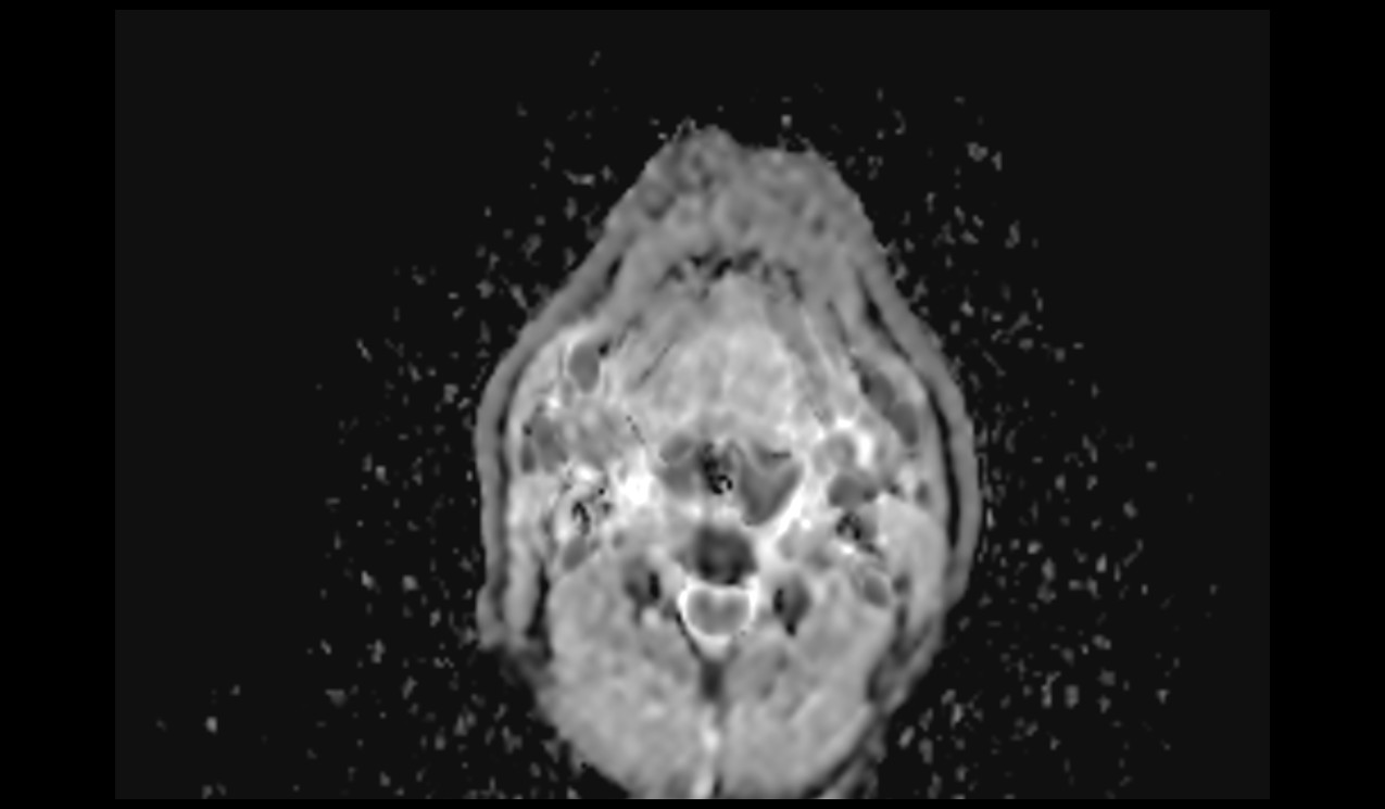
RESOLVE DWI Axial b800 Image of Neck

RESOLVE DWI Axial ADC Image of Neck
References
- Sheng, Y., Hong, R., Sha, Y., Zhang, Z., Zhou, K., & Fu, C. (2020). Performance of TGSE BLADE DWI compared with RESOLVE DWI in the diagnosis of cholesteatoma. BMC Medical Imaging, 20(Article number: 40), 3786 Accesses, 18 Citations, 1 Altmetric. Published on 19 April 2020
- Qian, W., Li, Z., Chen, W., Yin, H., Zhang, J., Xu, J., & Hu, C. (2022). RESOLVE-DWI-based Deep Learning Nomogram for Prediction of Normal-sized Lymph Node Metastasis in Cervical Cancer: A Preliminary Study. BMC Medical Imaging, 22(1), 221. doi:10.1186/s12880-022-00948-6.
- Tullos, H., Dale, B., Bidwell, G., Perkins, E., Raucher, D., Khan, M., & James, J. (2012). Multi-Shot RESOLVE Compared to Single-Shot EPI Diffusion-Weighted MR Imaging Acquisition Scheme. Medical Physics, 39(6Part5), 3640.
- Porter, D. A., & Heidemann, R. M. (2009). High resolution diffusion‐weighted imaging using readout‐segmented echo‐planar imaging, parallel imaging and a two‐dimensional navigator‐based reacquisition. Magnetic Resonance in Medicine, 62(2), 468-475.
- Morelli, J. N., Runge, V. M., Ai, F., Attenberger, U., Vu, L., Schmeets, S. H., … & Kirsch, J. E. (2011). An image-based approach to understanding the physics of MR artifacts. Radiographics, 31(3), 849-866.
- Schultz, A., Schmitter, S., Trampel, R., & Turner, R. (2018). Design of readout-segmented echo-planar imaging at ultra-high fields with reduced power deposition. Magnetic Resonance in Medicine, 79(4), 2103-2112.