Phase encoding direction in MRI
The MRI phase encoding direction refers to one of the three spatial dimensions along which phase encoding gradients are applied during MRI image acquisition. It determines how spatial information is encoded into the MRI signal, crucial for reconstructing the spatial details of the imaged object. The phase encoding direction influences image resolution, susceptibility to artifacts, and scan duration, making its understanding essential for optimizing MRI protocols and image quality.
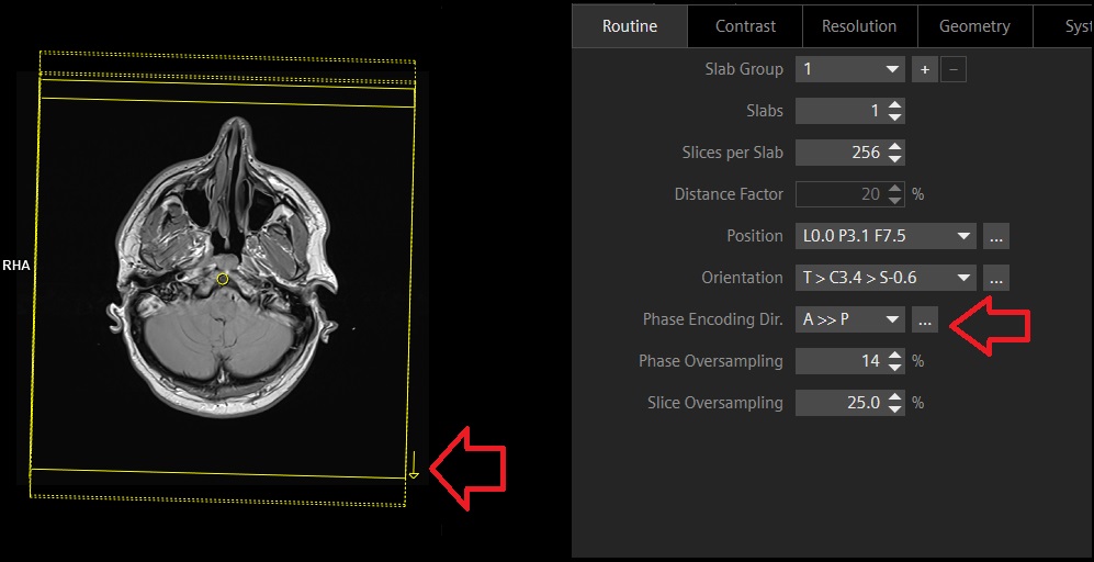
Picher displaying phase encoding direction choices in Siemens scanner configuration

Physics of Phase Encoding in MRI
MRI machines use strong magnetic fields and radio waves to create images of the body. The process involves several key steps:
Magnetic Field Application: A strong primary magnetic field, 𝐵0B0, aligns the protons in the body, predominantly those in water molecules, along its direction.
RF Pulse Excitation: A radiofrequency (RF) pulse is then applied perpendicular to 𝐵0B0, temporarily tipping the aligned protons away from the magnetic field direction.
Relaxation and Signal Emission: As the protons return to their original alignment with 𝐵0B0, they emit RF signals, which are detected and used to form images.
Spatial Encoding: To localize these signals spatially within the body, gradients in the magnetic field are used:
- Slice Selection Gradient: Selects the imaging slice.
- Frequency Encoding Gradient (Readout): Encodes spatial information along one axis (e.g., left-right or anterior-posterior).
- Phase Encoding Gradient: Encodes spatial information along another axis (e.g., anterior to posterior or right to left ). This is where the phase encoding direction comes in.
Phase Encoding Gradient: This gradient is applied in a specific direction (either x, y, or z-axis) perpendicular to the frequency encoding gradient. The phase encoding gradient momentarily alters the magnetic field strength across the scanned object in that direction, causing spins in different locations to accumulate phase at different rates. After this gradient is turned off, spins along the phase encoding direction will have accumulated different phase shifts based on their location. This variation in phase is used to spatially encode one dimension of the image.
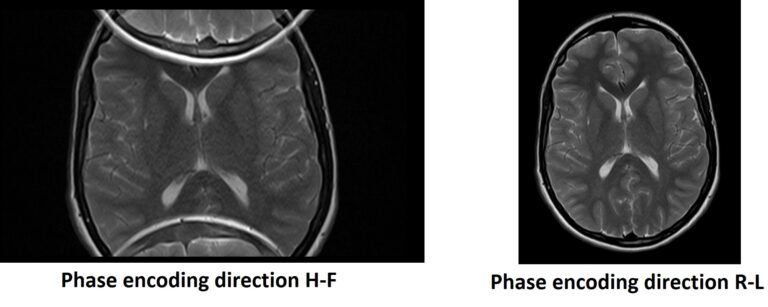
The image demonstrates the anterior to posterior phase encoding direction used in brain imaging.
The image demonstrates the right to left phase encoding direction used in abdomen imaging.

Importance of phase encoding in Scanning
Spatial Resolution: The direction and strength of the phase encoding gradient determine the resolution in the encoded direction. Adjusting the number of phase encoding steps can change the resolution.
Scan Time: The phase encoding process is repeated multiple times, once for each phase encoding step. Therefore, the number of steps in this direction is a key factor in determining the total scan time.
Artifact Reduction : Artifacts in MRI can occur due to various reasons including body motion, flow of fluids within the body, and machine errors. The phase encoding process can influence certain types of artifacts:
Artifacts Related to Phase Encoding Direction
- Motion Artifacts: Motion during the scan causes inconsistencies in the phase encoding steps, leading to ghosting or repeated patterns across the image. By adjusting the phase encoding direction, the impact of motion (such as breathing ) can be minimized. For instance, setting the phase encoding direction perpendicular to the main direction of motion can reduce these effects.
- Aliasing (Wrap-around) Artifacts: If the body part being imaged extends beyond the field of view in the phase encoding direction, it can lead to aliasing, where signals from outside the field of view wrap around to the opposite side of the image. By appropriately setting the phase encoding direction and adjusting the field of view, these artifacts can be reduced.
Flow Artifacts : Flow artifacts are particularly prevalent in areas with significant movement of fluids, such as blood in arteries and veins. These artifacts manifest as distortions or signal voids in the images. Changing the phase encoding direction can move these artifacts away from the area of interest. This technique is especially useful in cardiac imaging, where blood flow can significantly affect image quality.
Elimination of Aliasing or Wrap-around Artifact in MRI through Phase Swapping
Limitations in Phase Encoding Directions
In MRI scans, the phase encoding direction is usually set orthogonal to the frequency encoding direction. The common phase encoding directions are limited based on the orientation of the scan:
- Axial Scans: Phase encoding is typically performed in the anterior-to-posterior (A-P) or right-to-left (R-L) direction.
- Coronal Scans: The common phase encoding directions are right-to-left (R-L) and head-to-foot (H-F).
- Sagittal Scans: Typical directions include anterior-to-posterior (A-P) and head-to-foot (H-F).