MRI Sports Hernia Protocol
What is Sports Hernia?
A sports hernia, also known as athletic pubalgia or inguinal disruption, is a painful condition that affects the groin area, typically occurring in athletes involved in sports that require sudden changes in direction or intense twisting movements. Despite its name, a sports hernia is not a true hernia involving a protrusion of abdominal organs through a weakened muscle or tissue. Instead, it is a complex soft tissue injury involving the muscles, tendons, and ligaments of the lower abdomen and groin.
The exact cause of a sports hernia is not well understood, but it is believed to result from repetitive stress or strain on the muscles and tendons in the groin area. This can lead to micro-tears and inflammation, causing pain and discomfort. Sports hernias often occur in athletes participating in sports like soccer, ice hockey, rugby, tennis, and football.
The symptoms of a sports hernia typically include groin pain that worsens with physical activity, especially during movements that involve twisting, turning, or kicking. The pain may be localized or radiate to the lower abdomen, adductor muscles, or testicles. Unlike a traditional hernia, there is usually no visible bulge or protrusion in the affected area.
In the context of sports hernias, an MRI scan is a valuable tool for assessing and determining the magnitude of soft tissue injuries occurring in the groin and lower abdominal area. It enables the visualization of various structures, including muscles, tendons, ligaments, and other relevant components implicated in this condition. By utilizing this imaging technique, healthcare professionals can effectively distinguish sports hernias from alternative sources of groin pain, such as hernias, muscle strains, or issues related to the hip joint.
Indications for MRI Sports Hernia scan
- Suspicion of a rectus abdominis injury, adductor injury and osteitis pubis,
- groin pain in athletes
- Athletic pubalgia
Contraindications
- Any electrically, magnetically or mechanically activated implant (e.g. cardiac pacemaker, insulin pump biostimulator, neurostimulator, cochlear implant, and hearing aids)
- Intracranial aneurysm clips (unless made of titanium)
- Pregnancy (risk vs benefit ratio to be assessed)
- Ferromagnetic surgical clips or staples
- Metallic foreign body in the eye
- Metal shrapnel or bullet
Patient preparation for MRI Sports Hernia scan
- A satisfactory written consent form must be taken from the patient before entering the scanner room
- Ask the patient to remove all metal objects including keys, coins, wallet, cards with magnetic strips, jewellery, hearing aid and hairpins
- Ask the patient to undress and change into a hospital gown
- If possible provide a chaperone for claustrophobic patients (e.g. relative or staff )
- Offer earplugs or headphones, possibly with music for extra comfort
- Explain the procedure to the patient
- Instruct the patient to keep still
- Note the hight and weight of the patient
Positioning for Sports Hernia MRI
- Position the patient in supine position with head pointing towards the magnet (head first supine)
- Position the patient over the spine coil and place the body coil over the pelvis ( iliac crest down to mid thigh)
- Securely tighten the body coil using straps to prevent respiratory artefacts
- Give a pillow under the head for extra comfort
- Centre the laser beam localiser over symphysis pubis (4 inches below iliac crest)

Recommended MRI Sports Hernia Protocols and Planning
localiser
To localize and plan the sequences, it is essential to initially acquire a three-plane T2 HASTE localizer. These fast single-shot localizers have an acquisition time of under 25 seconds and are highly effective in accurately localizing pelvic structures.

T2 tse DIXON (T2+T2 FAT SAT) axial 3mm
Plan the axial slices on the coronal plane, angling the positioning block parallel to the right and left hip. Verify the positioning block in the other two planes and ensure an appropriate angle is applied in the sagittal plane, which should be horizontally across the rectus abdominal muscle. The slices should adequately cover the lower abdomin from 2 inches above the acetabulam down to 2inches below the pubic sypisis. To minimize ghosting artifacts caused by peristalsis and breathing, consider using a saturation band over the axial block.

Parameters
TR 5000-6000 | TE 110 | FLIP 150 | NEX 2 | SLICE 3 MM | MATRIX 320X320 | FOV 300-330 | PHASE R>L | GAP 10% | FAT SAT DIXON |
T2 tse DIXON (T2+T2 FAT SAT) sagittal 3mm
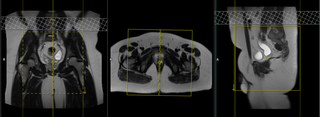
Plan the sagittal slices on the coronal plane and angle the positioning block parallel to the pubic symphysis. Verify the positioning block in the other two planes. Provide an appropriate angle in the axial plane, parallel to the line connecting the pubic symphysis and the anal canal. The slices should adequately cover both groins, extending from the right acetabulum to the left acetabulum. To minimize ghosting artifacts caused by peristalsis and breathing, consider using a saturation band over the sagittal block.
Parameters
TR 5000-6000 | TE 90-120 | SLICE 3 MM | FLIP 151 | PHASE H>F | MATRIX 320X320 | FOV 200-250 | GAP 10% | NEX(AVRAGE) 2 |
T2 tse DIXON(T2+T2 FAT SAT) coronal 3mm
Plan the coronal slices on the sagittal plane and angle the positioning block vertically across the abdomen. Verify the positioning block in the other two planes and provide an appropriate angle in the axial plane, aligning it parallel to the right and left ischial tuberosity. The slices should adequately cover the lower abdomen, extending from 1 inch anterior to the symphysis pubis to the ischial tuberosity. To minimize ghosting artifacts caused by peristalsis and breathing, consider using a saturation band over the coronal block.

Parameters
TR 5000-6000 | TE 90-120 | SLICE 3 MM | FLIP 151 | PHASE H>F | MATRIX 320X320 | FOV 250-300 | GAP 10% | NEX(AVRAGE) 2 |
T1 tse coronal 3mm
Plan the coronal slices on the sagittal plane and angle the positioning block vertically across the abdomen. Verify the positioning block in the other two planes and provide an appropriate angle in the axial plane, aligning it parallel to the right and left ischial tuberosity. The slices should adequately cover the lower abdomen, extending from 1 inch anterior to the symphysis pubis to the ischial tuberosity. To minimize ghosting artifacts caused by peristalsis and breathing, consider using a saturation band over the coronal block.
Parameters
TR 400-600 | TE 15-25 | SLICE 3 MM | FLIP 140 | PHASE R>L | MATRIX 384X320 | FOV 250-300 | GAP 10% | NEX(AVRAGE) 2 |
T2 tse DIXON(T2+T2 FAT SAT) coronal oblique 3mm
Plan the coronal slices on the sagittal plane, angling the positioning block parallel to the anterior iliac crest. Verify the positioning block in the other two planes and ensure an appropriate angle in the axial plane, aligning it parallel to the right and left ischial tuberosity. The slices should adequately cover the lower abdomen, extending from 2 inches anterior to the symphysis pubis to the ischial tuberosity. To minimize ghosting artifacts caused by peristalsis and breathing, consider using a saturation band over the coronal block.

Parameters
TR 5000-6000 | TE 110 | FLIP 150 | NEX 2 | SLICE 3 MM | MATRIX 256X256 | FOV 250-300 | PHASE R>L | GAP 10% | FAT SAT DIXON |