MRI FABS View : Protocol and Planning
Indications for FABS View MRI
- Distal biceps brachii tendon injury
Contraindications
- Any electrically, magnetically or mechanically activated implant (e.g. cardiac pacemaker, insulin pump biostimulator, neurostimulator, cochlear implant, and hearing aids)
- Intracranial aneurysm clips (unless made of titanium)
- Pregnancy (risk vs benefit ratio to be assessed)
- Ferromagnetic surgical clips or staples
- Metallic foreign body in the eye
- Metal shrapnel or bullet
Patient preparation for FABS View MRI
- A satisfactory written consent form must be taken from the patient before entering the scanner room
- Ask the patient to remove all metal objects including keys, coins, wallet, cards with magnetic strips, jewellery, hearing aid and hairpins
- If possible provide a chaperone for claustrophobic patients (e.g. relative or staff )
- Offer earplugs or headphones, possibly with music for extra comfort
- Explain the procedure to the patient
- Instruct the patient to keep still
- Note the hight and weight of the patient
Positioning for FABS View MRI
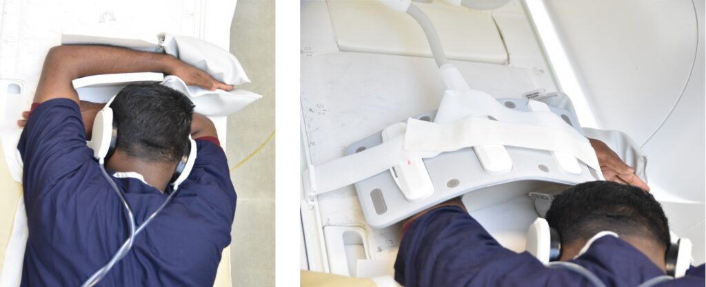
- Positioning the patient prone over the spine coil, with the elbow flexed, shoulder abducted, and forearm supinated.
- Immobilize the elbow and forearm with sandbags and cushions.
- Position the flex coil horizontally across the elbow joint.
- Centre the laser beam localiser over the elbow joint
- Register the patient on the scanner as 'head first supine'
Recommended FABS View MRI Protocols and Planning
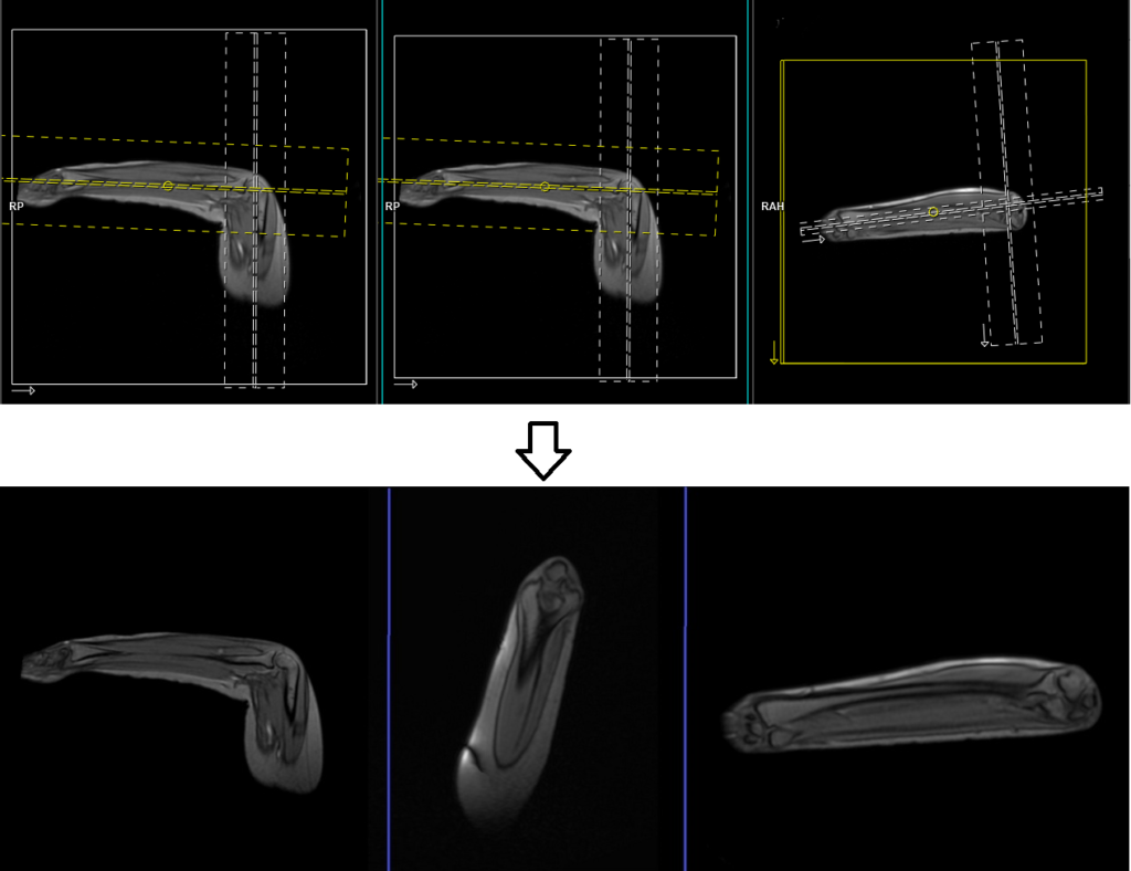
localiser
A two-plane large FOV localizer must be taken at the beginning to localize and plan the sequences. Typically, these localizers take less than 25 seconds and can be achieved using T1 weighted low-resolution scans. It is advisable to obtain additional localizers until you have acquired accurate axial, coronal, and sagittal localizer images.

localiser 2
For proper planning of the FABS view, it is necessary to include a second three-plane localizer. The first two localizers should be planned in the sagittal plane. Align the first planning block parallel to the humerus for the coronal plane, and align the second planning block parallel to the radius and ulna for the axial localizer. The last localizer should be planned based on the initial coronal localizer of the forearm and aligned parallel to the radius and ulna to obtain a proper sagittal localizer.
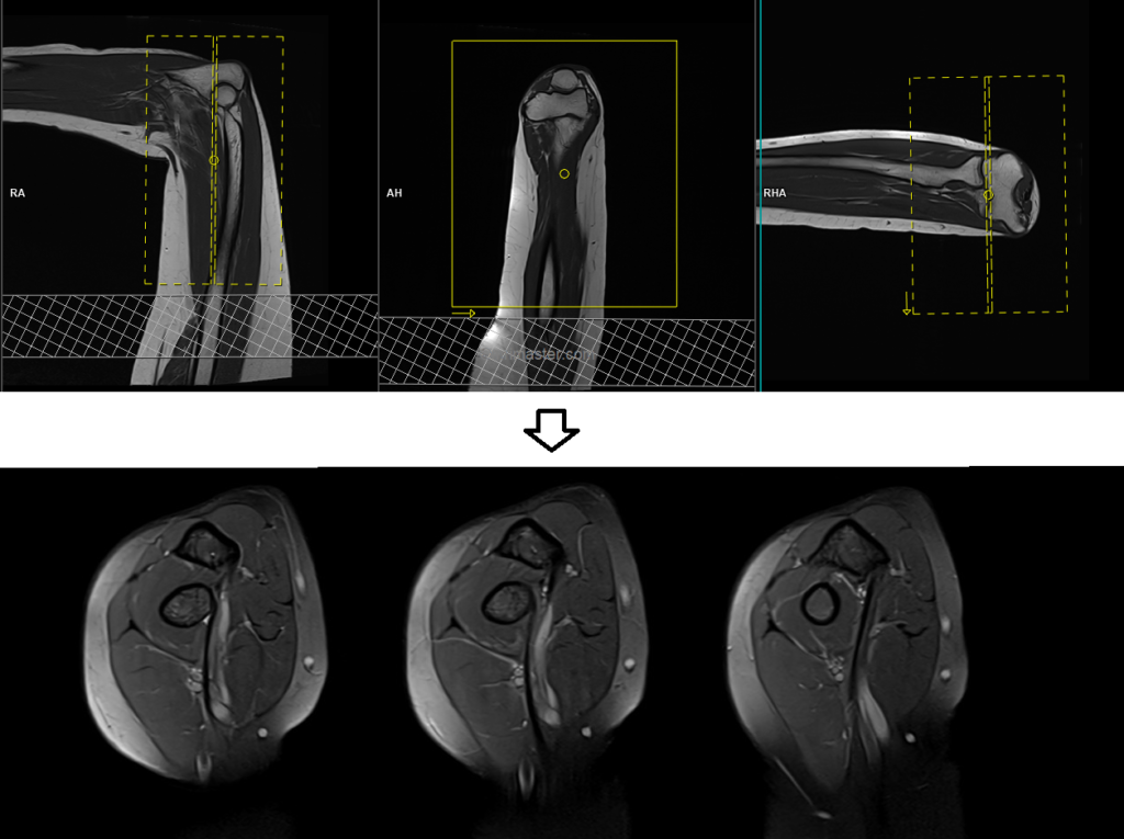
PD Fat saturated coronal 2mm SFOV
Plan the coronal slices on the sagittal plane and angle the planning block parallel to the humerus. Check the positioning block in the other two planes. An appropriate angle must be used in the axial plane (perpendicular to the radius and ulna or parallel to the medial and lateral epicondyle of the humerus). Slices must be sufficient to cover the entire humerus from the radial tuberosity to the triceps brachii tendon. Adding saturation bands to the bottom of the coronal block will help reduce arterial pulsation artifacts.
Parameters
TR 3000-4000 | TE 15-20 | SLICE 2 MM | FAT SAT ON | PHASE A>P | MATRIX 320X256 | FOV 130-170 | GAP 10% | NEX(AVRAGE) 2 |
T1 tse coronal 2mm SFOV
Plan the coronal slices on the axial plane and angle the positioning block parallel to the medial and lateral humeral epicondyles. Check the positioning block in the other two planes. An appropriate angle must be used in the sagittal plane (parallel to the humerus and ulna). Slices must be sufficient to cover the whole elbow joint from the anterior to posterior aspect. Adding saturation bands on the top and bottom of the coronal block will help to reduce arterial pulsation artifacts. The phase direction must be head to feet with a minimum of 100% oversampling to avoid wrap-around and pulsation artifacts.

Parameters
TR 400-500 | TE 15-20 | FLIP 150 | NEX 2 | SLICE 2 MM | MATRIX 320X256 | FOV 130-170 | PHASE A>P | GAP 10% | oversample 0% |